
Meet the Expert
Identification and history
Name: Artù
Report and medical history: horse, Paint Horse, male, 5 years old.
Following a long journey, the subject began to manifest cough, dyspnea, and fever
Diagnostics
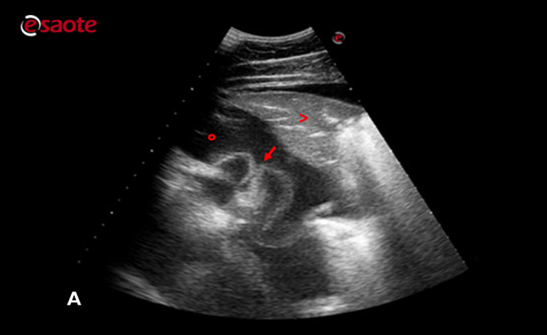 Image A
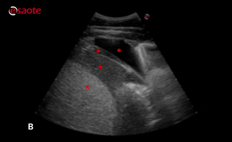 Image B
Echocardiographic images were acquired using the MyLab™OmegaVET system.
Ultrasound examination of the left side of the thorax revealed a moderate amount of anechoic fluid (o) in the pleural cavity (Image A). There are also large areas of consolidated lung (>). In image A, the presence of the fluid allows us to visualize the phrenico-pericardial ligament (arrow) as a filiform hyperechogenic structure floating in it. In image B, the diaphragm (*), liver (+), and spleen (<) can also be identified.
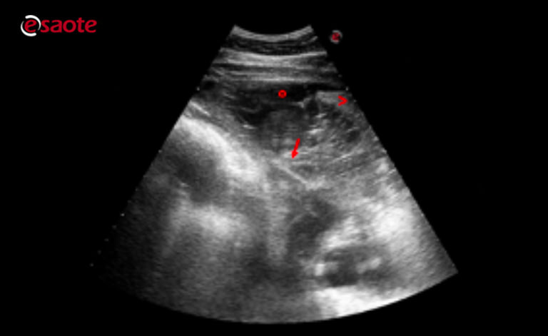 Image C
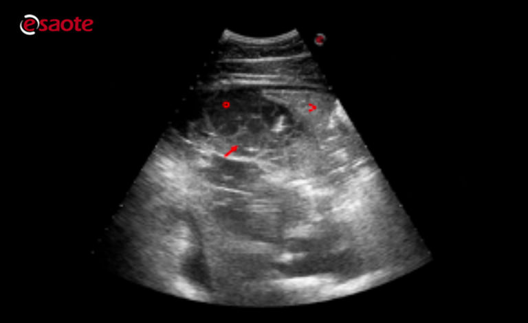 Image D
Echocardiographic images were acquired using the MyLab™OmegaVET system.
At the right side of the thorax, the ultrasound images show areas of consolidated lung (>) and a moderate amount of pleural fluid (o) in which the presence of fibrin (arrow) is particularly evident, organizing into a dense network and multiple concamerations.
Further examinations: an endoscopic examination of the airways revealed the presence of pharyngeal lymphoid hyperplasia and a significant presence of mucopurulent exudate in the tracheal lumen. The culture test performed on the tracheal aspirate fluid revealed the presence of enrofloxacin sensitive Streptococcus spp.
Conclusions and treatment:
Equine Medicine Unit,
Università degli studi di Milano, Ospedale Universitario Veterinario di Lodi
Severe bacterial pleuropneumonia
The patient was admitted to intensive care and received antibiotic and anti-inflammatory treatment, combined with ultrasound monitoring of the chest and drainage of the pleural effusion. Gastroprotectants and probiotics were also administered.
MyLab is a trademark of Esaote spa.
Product images are for illustrative purposes only. For further details, please contact your Esaote sales representative.
Technology and features are system/configuration dependent. Specifications subject to change without notice. Information might refer to products or modalities not yet approved in all countries.
Read other VET interviews

Feline Ultrasonography
Report and medical history: cat, Common European, spayed female, 16 years old.
The patient presented at our clinic with dejection, anorexia, hematochezia and vomiting. On clinical examination there was depressed sensory status ...

Canine Echocardiography
Report and medical history: dog, dachshund, female, 13 years old.
An abdominal ultrasound and echocardiogram was requested for loss of appetite, lethargy, and syncopal episodes ...

Canine Ultrasonography
Dog, Short-haired Dachshund, Female, 10 years old
Ultrasound checkup required for weight loss, PU/PD, recurrent vomiting, especially after ingestion of extra-diet foods ...