Ultrasound Clinical Images
Just like space, some body parts are not visible to the naked eye: that’s why we need Esaote’s advanced diagnostic imaging technologies to be able to explore them and unlock their secrets.
Outstanding Ultrasound clinical images highlight the Esaote's focus in delivering innovative clinical solutions from prevention to therapeutic applications: a wide clinical gallery with high-res image quality means added diagnostic value and makes a difference for both clinicians and patients.
Today Esaote supports healthcare professionals with diagnostic imaging systems to deliver accuracy and precision of the clinical image as required by doctors on one hand, quality of diagnosis to improve the quality of life of the patients on the other and finally making what is essential visible to the eye and contributing to build a better future for everyone.
Filter by application
Filter by product
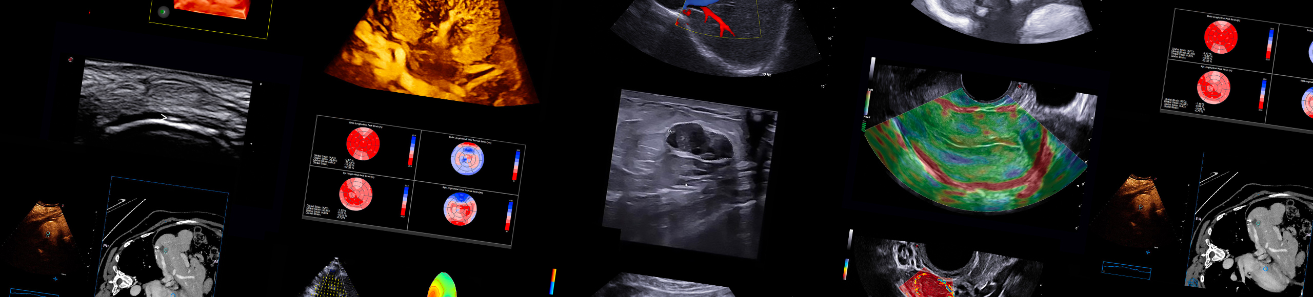
MyLab™E85 GTS Edition - Biopsy guide’s simultaneous display
MyLab™E85 GTS Edition - Biopsy guide’s simultaneous display
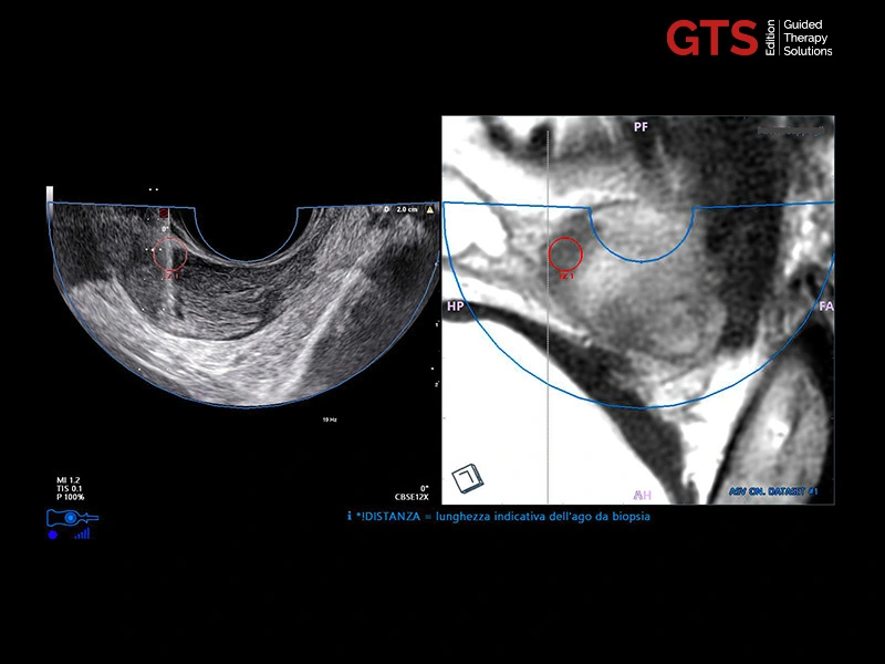
MyLab™E85 GTS Edition - Prostate segmentation for systematic biopsies
MyLab™E85 GTS Edition - Prostate segmentation for systematic biopsies
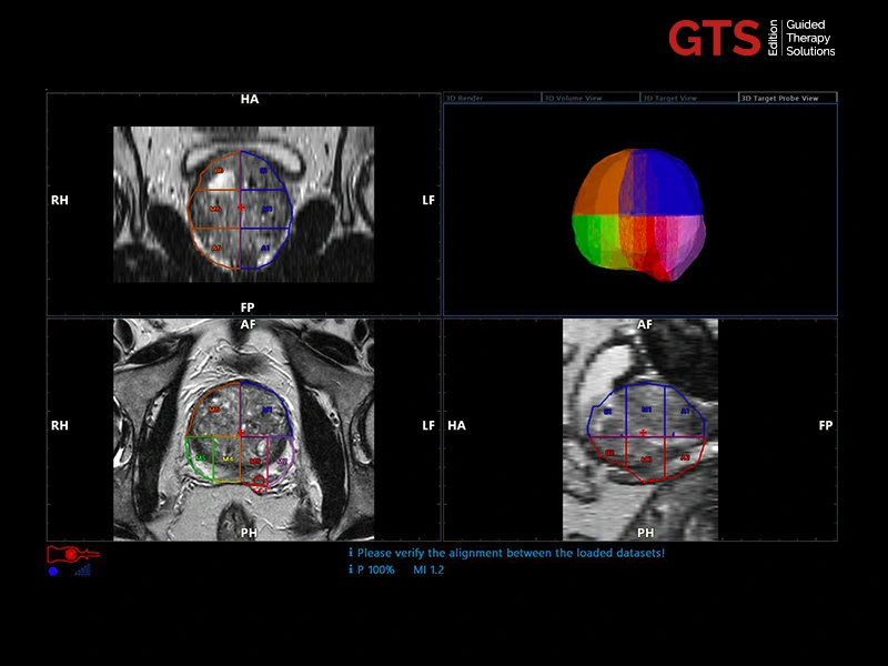
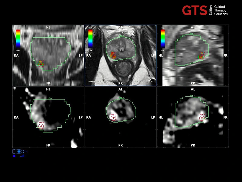
MyLab™E85 GTS Edition - Automated prostate report
MyLab™E85 GTS Edition - Automated prostate report
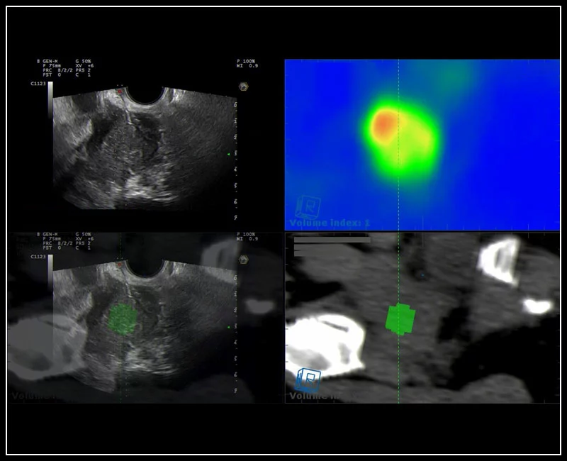
MyLab™E85 GTS Edition - Prostate Attention Map
MyLab™E85 GTS Edition - Prostate Attention Map
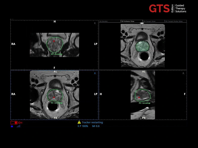
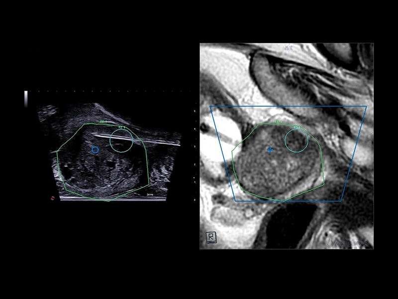
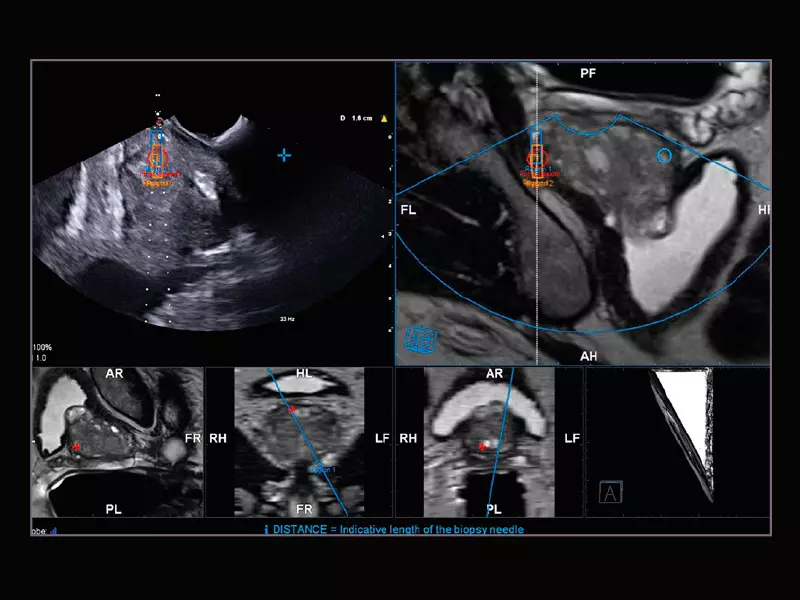
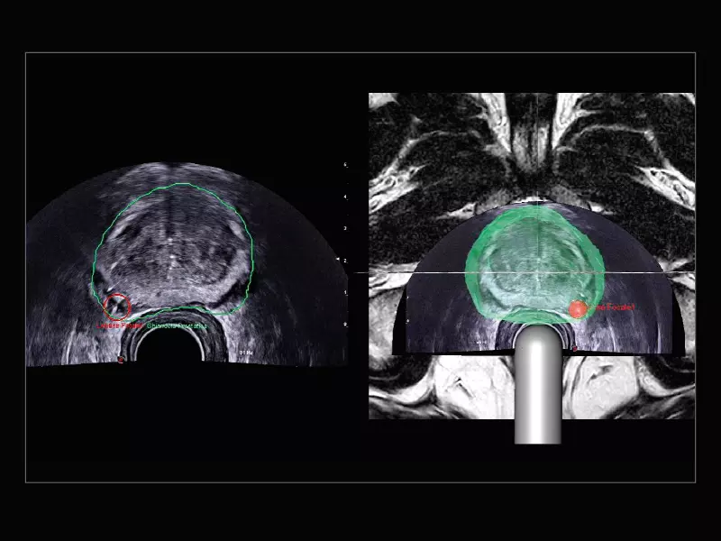
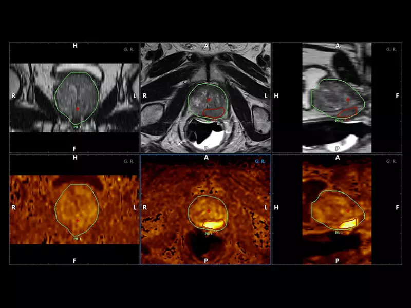
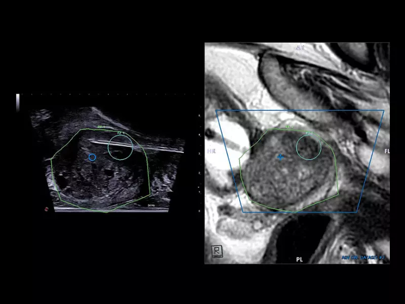
MyLab™E85 GTS Edition - MRI prostate AI-based segmentation
MyLab™E85 GTS Edition - MRI prostate AI-based segmentation
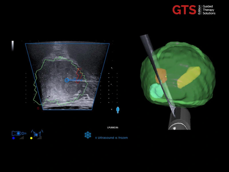
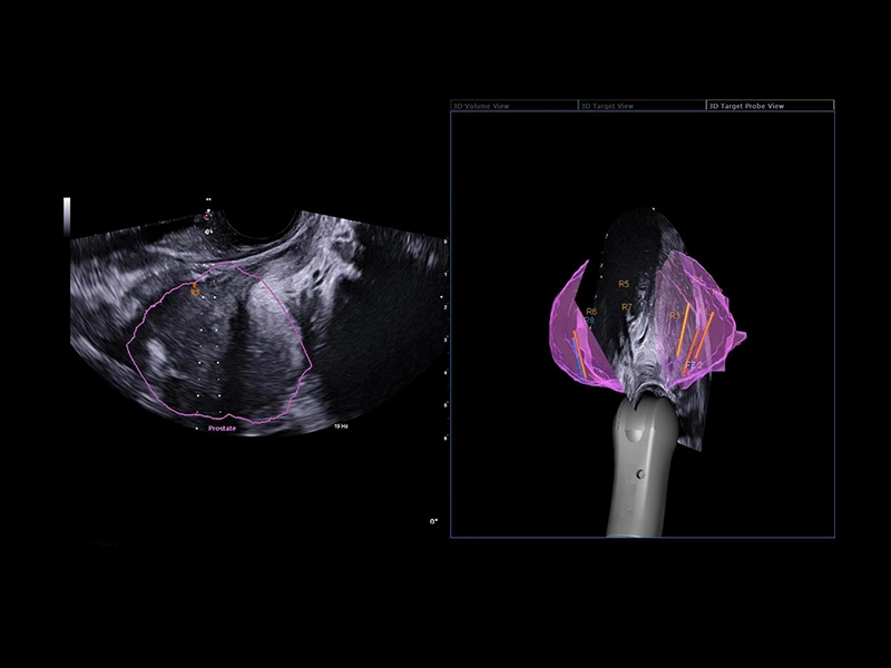
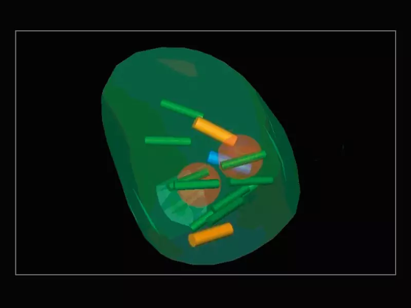
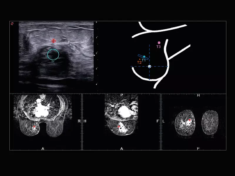
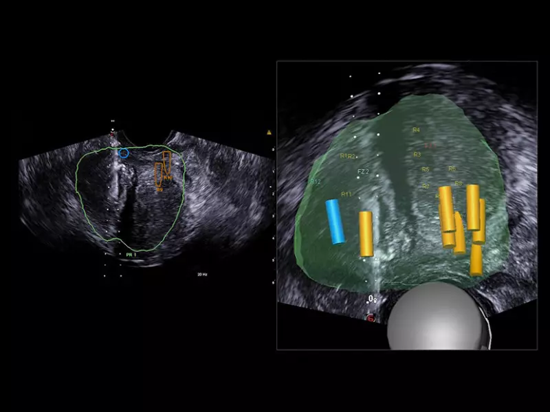
MyLab™E85 GTS Edition - Navigation environment with real-time 3D model of prostate (targets and biopsy cores)
MyLab™E85 GTS Edition - Navigation environment with real-time 3D model of prostate (targets and biopsy cores)
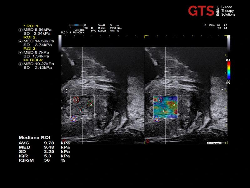
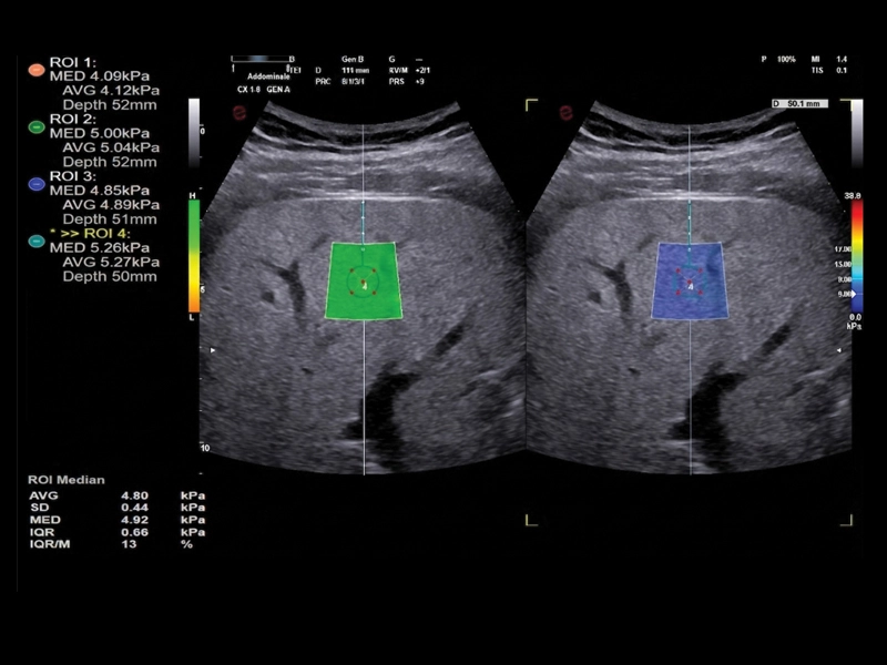
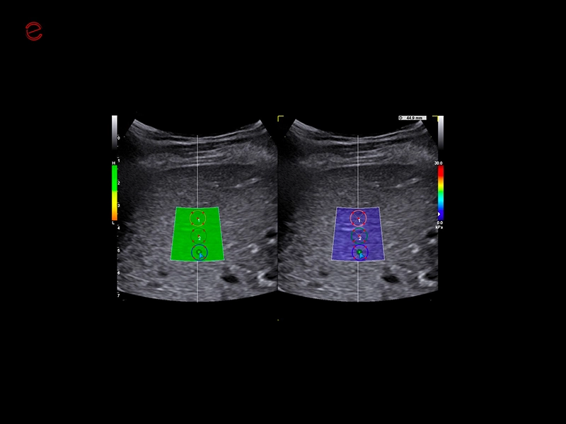
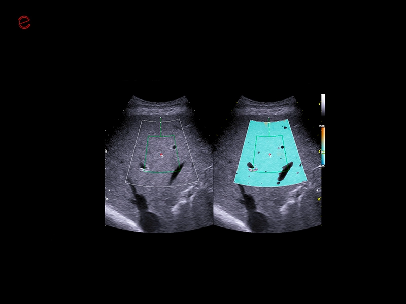
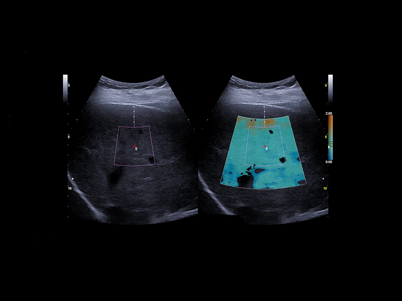
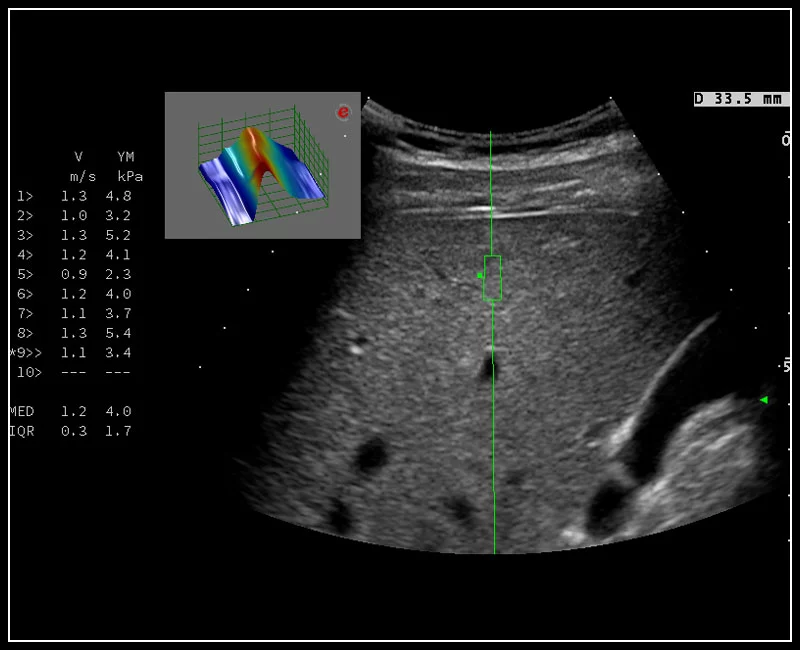
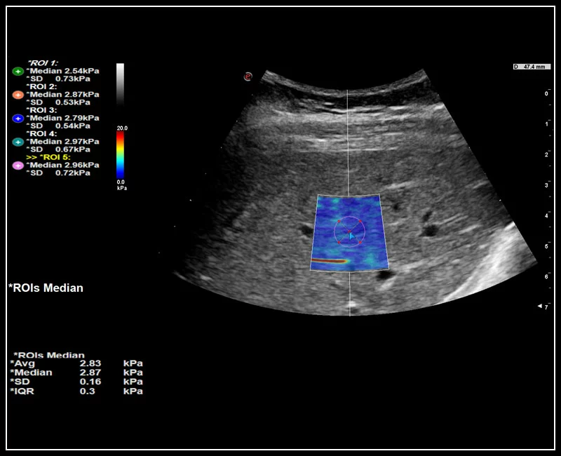
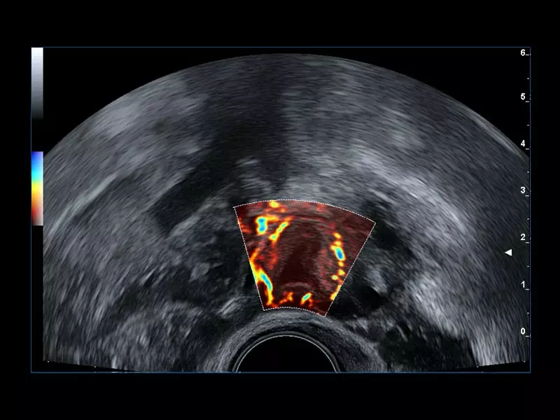
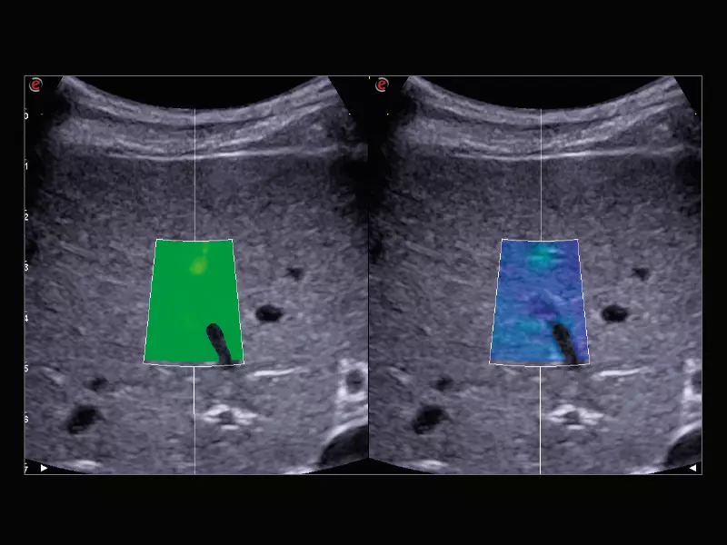
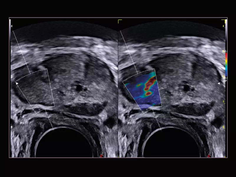
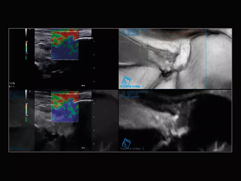
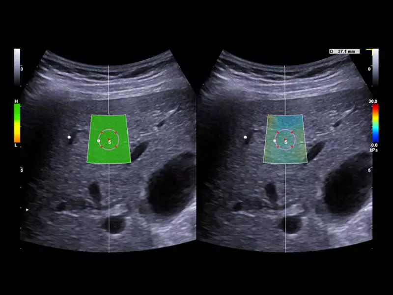
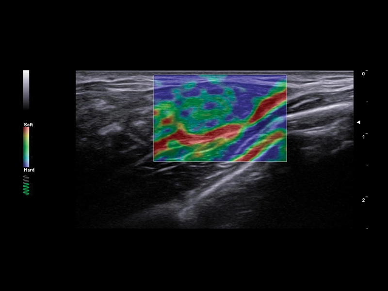
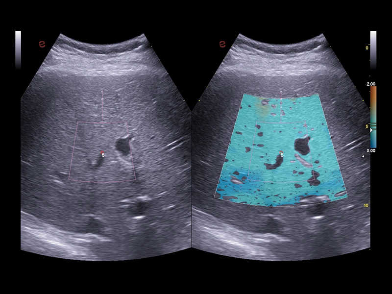
MyLab™E85 GTS Edition - Shear-Wave elastography technology in prostate
MyLab™E85 GTS Edition - Shear-Wave elastography technology in prostate
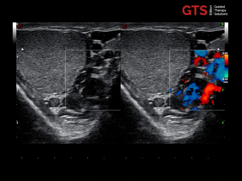
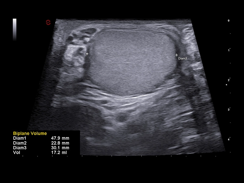
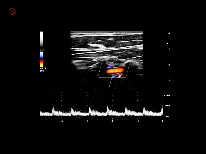
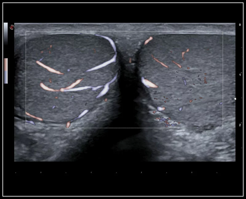
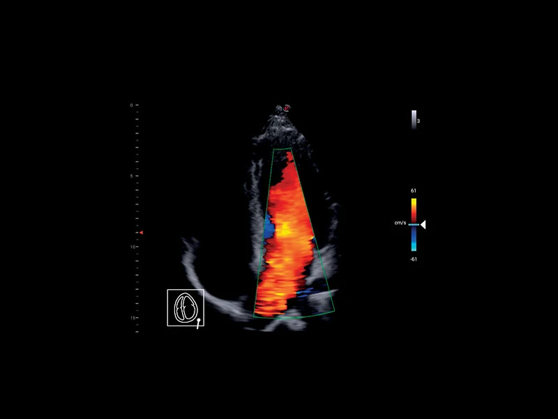
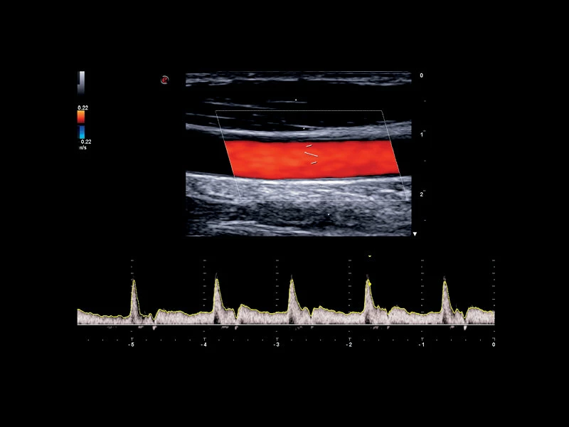
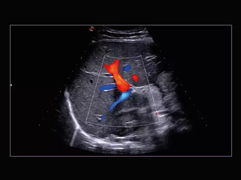
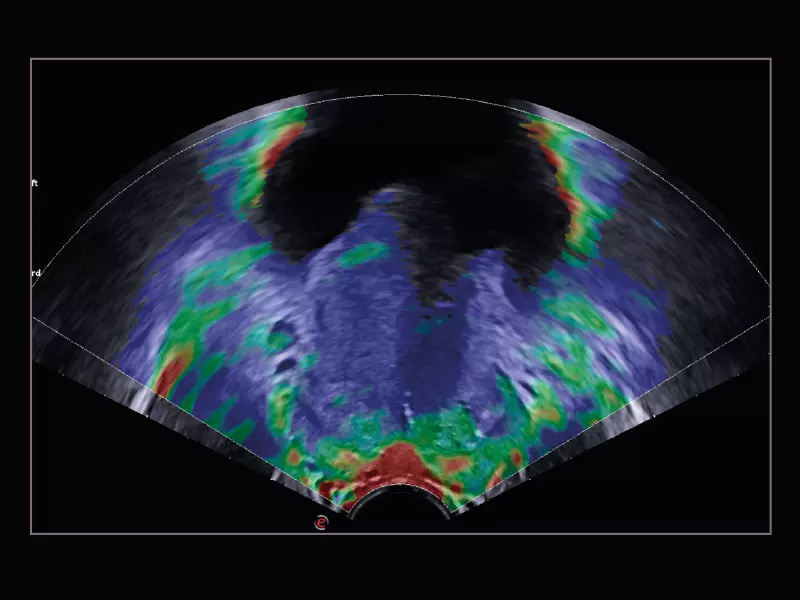
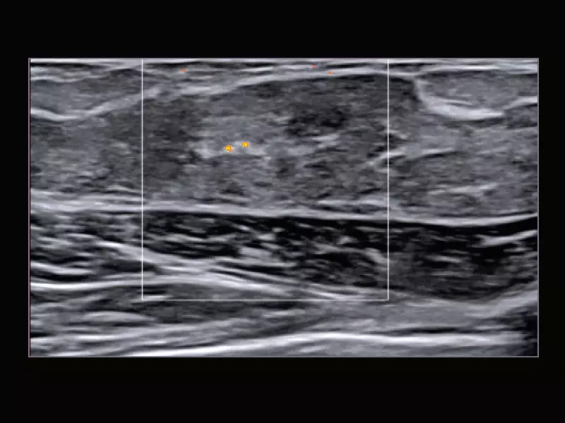
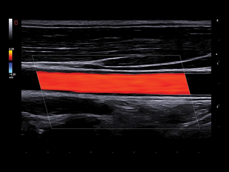
MyLab™E85 GTS Edition - Testis with Color Doppler technology
MyLab™E85 GTS Edition - Testis with Color Doppler technology
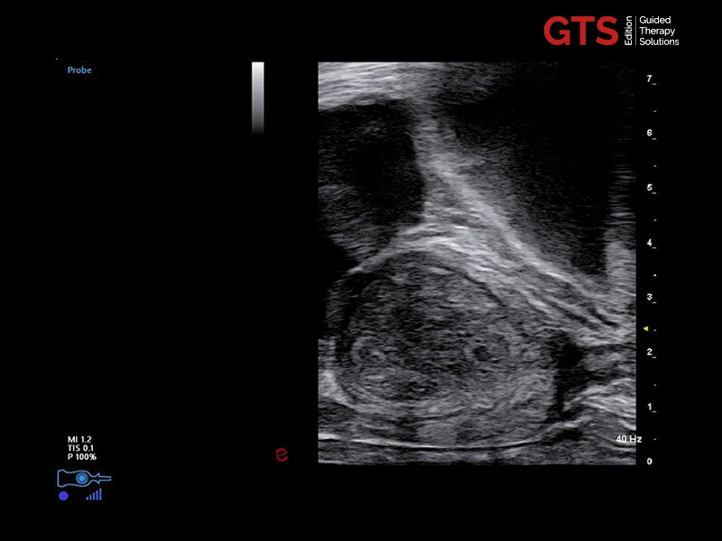
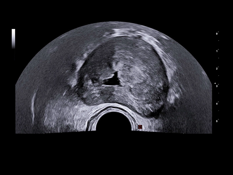
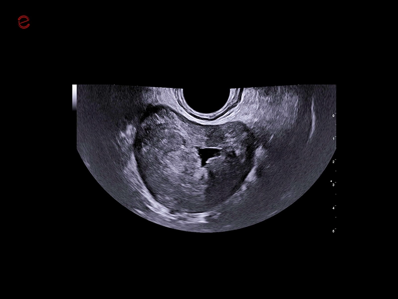
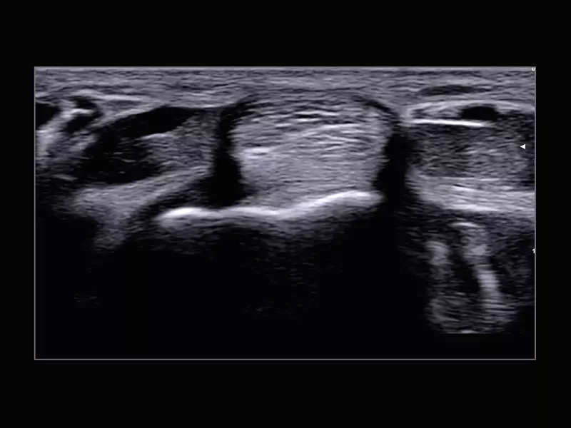
MyLab™E85 GTS Edition - Prostate image in transperineal biopsy approach
MyLab™E85 GTS Edition - Prostate image in transperineal biopsy approach
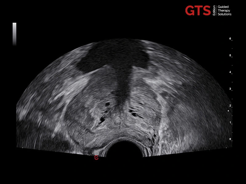
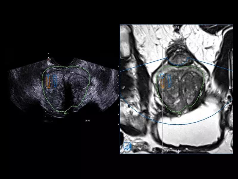
MyLab™E85 GTS Edition - Prostate image in transrectal biopsy approach
MyLab™E85 GTS Edition - Prostate image in transrectal biopsy approach
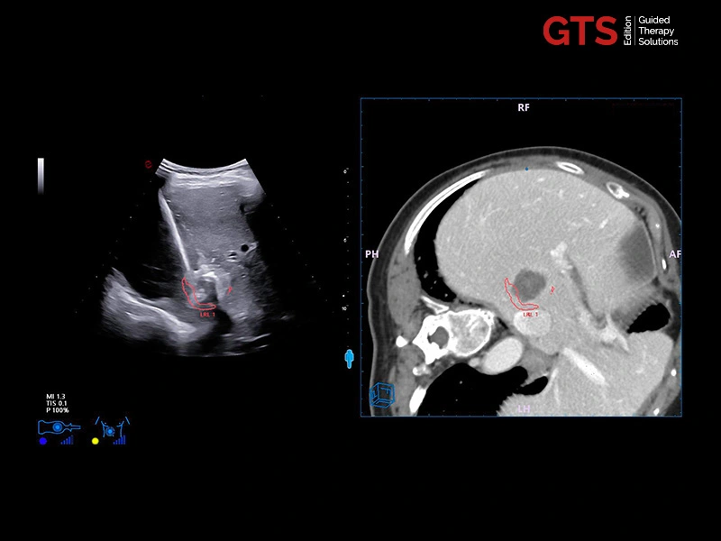
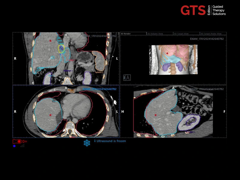
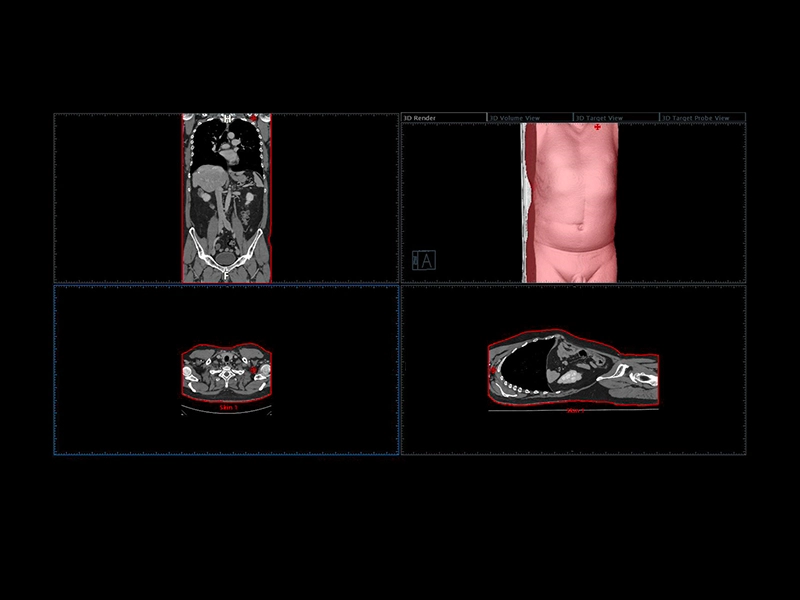
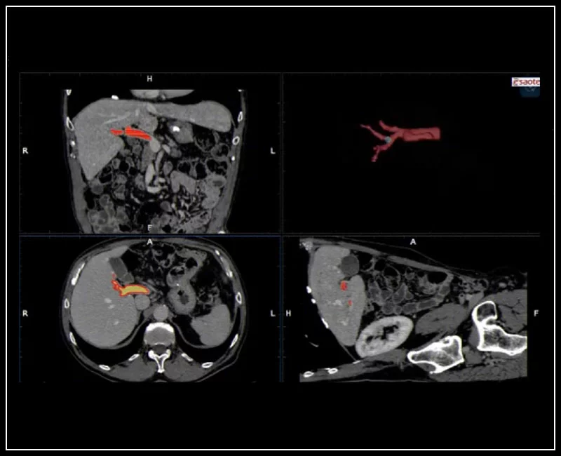
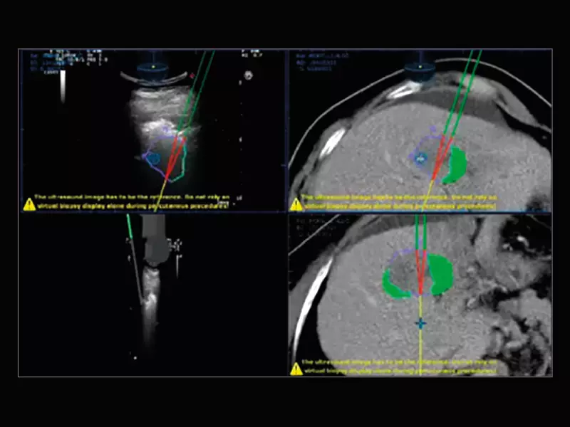
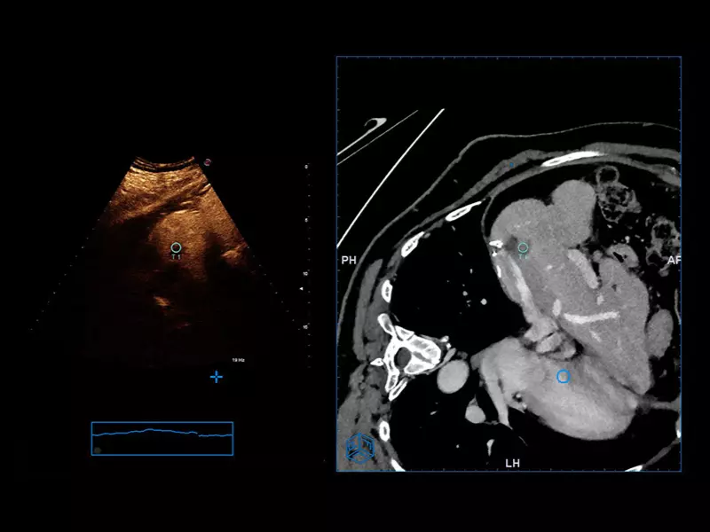
MyLab™E85 GTS Edition - Liver fusion - Residual zone as a new target on CT-imaging data
MyLab™E85 GTS Edition - Liver fusion - Residual zone as a new target on CT-imaging data
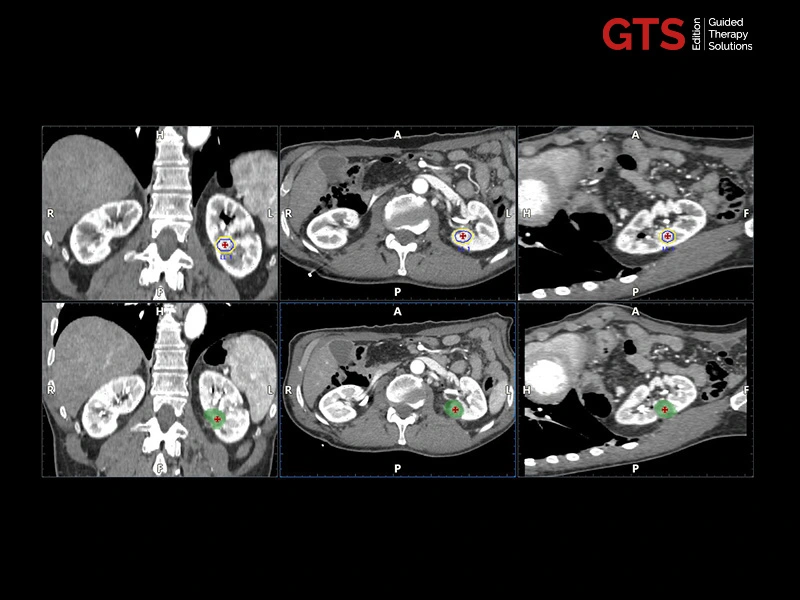
MyLab™E85 GTS Edition - Kidney fusion – Pre/post-ablation CT data comparison
MyLab™E85 GTS Edition - Kidney fusion – Pre/post-ablation CT data comparison
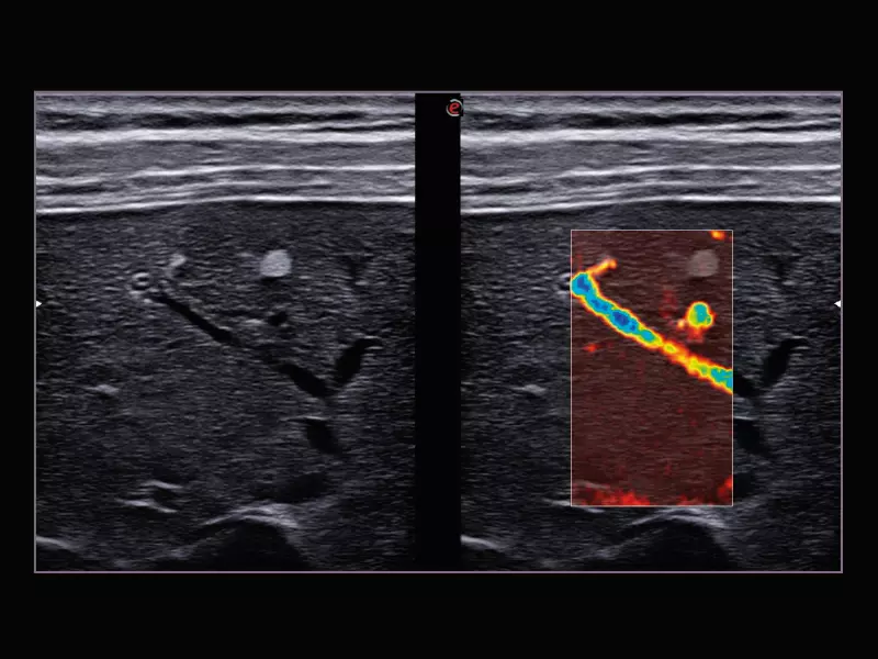
MyLab™E85 GTS Edition - Needle tracking combined with CnTITM Clear mode
MyLab™E85 GTS Edition - Needle tracking combined with CnTITM Clear mode
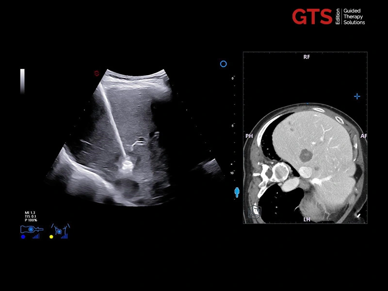
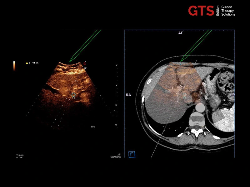
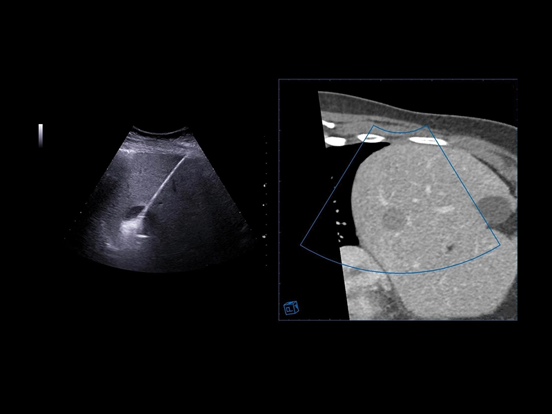
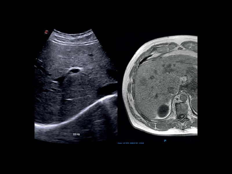
MyLab™E85 GTS Edition - Liver fusion imaging between CT and real-time US
MyLab™E85 GTS Edition - Liver fusion imaging between CT and real-time US
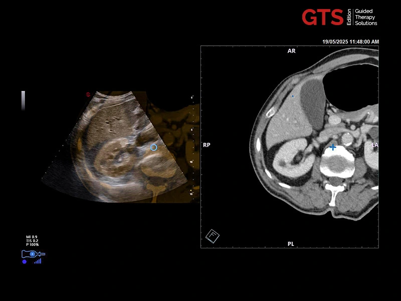
MyLab™E85 GTS Edition - Kidney fusion imaging between CT and real-time US
MyLab™E85 GTS Edition - Kidney fusion imaging between CT and real-time US
MyLab™E85 GTS Edition - AI-based organ segmentation tool
MyLab™E85 GTS Edition - AI-based organ segmentation tool
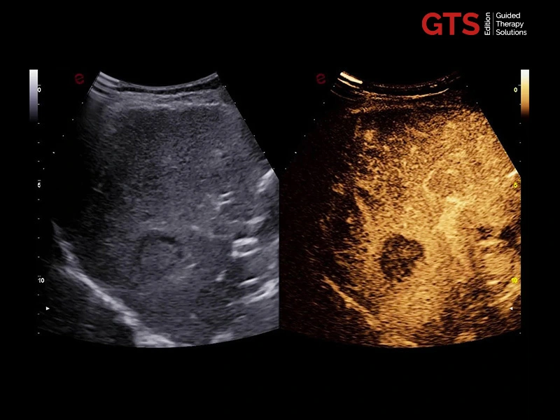
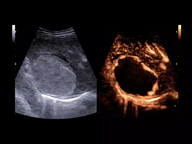
MyLab™E85 GTS Edition - LIver CnTITM Clear with linear probe
MyLab™E85 GTS Edition - LIver CnTITM Clear with linear probe
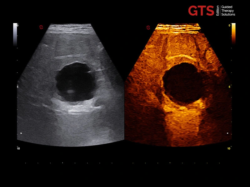
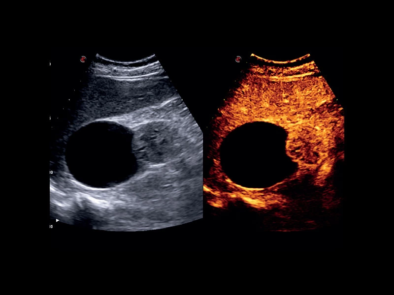
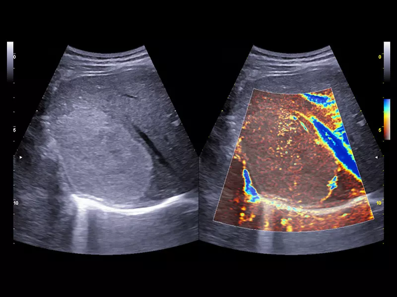
MyLab™E85 GTS Edition - Kidney CnTITM Clear with convex probe
MyLab™E85 GTS Edition - Kidney CnTITM Clear with convex probe
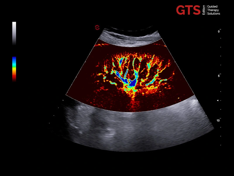
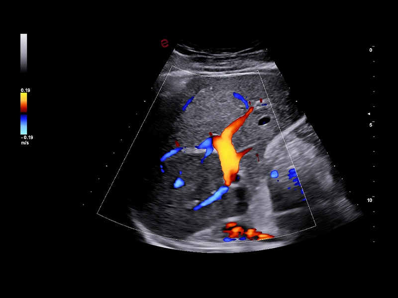
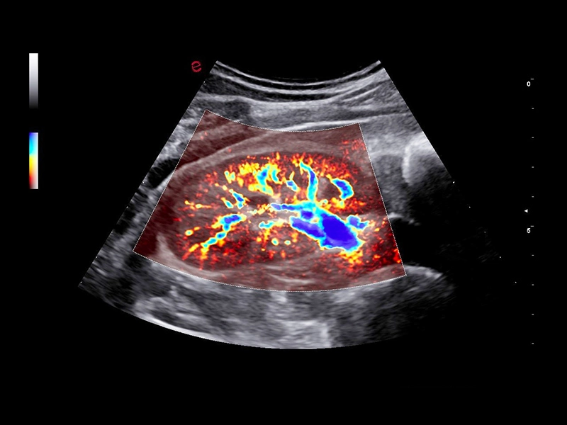
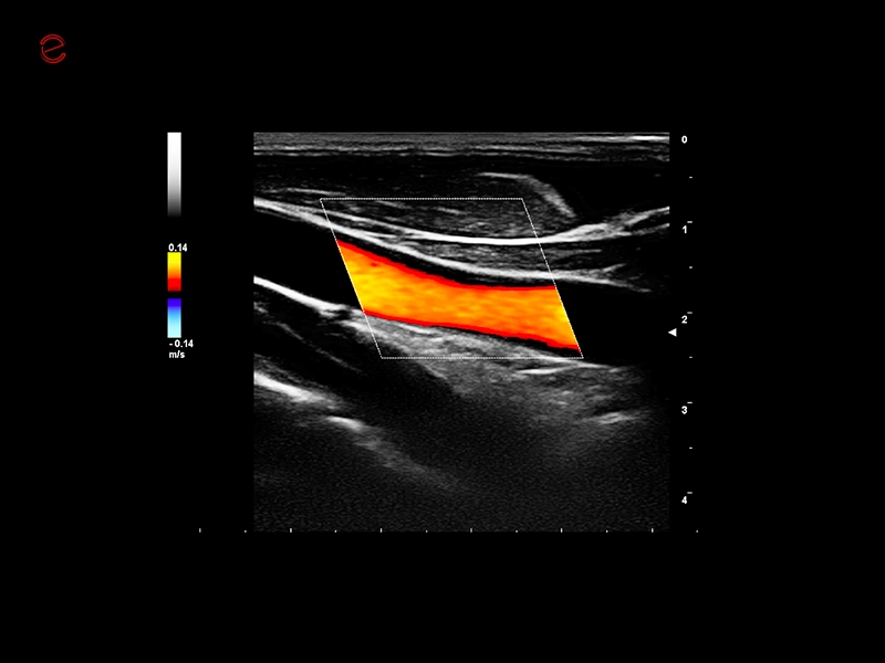
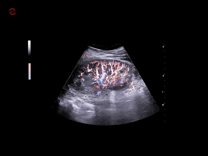
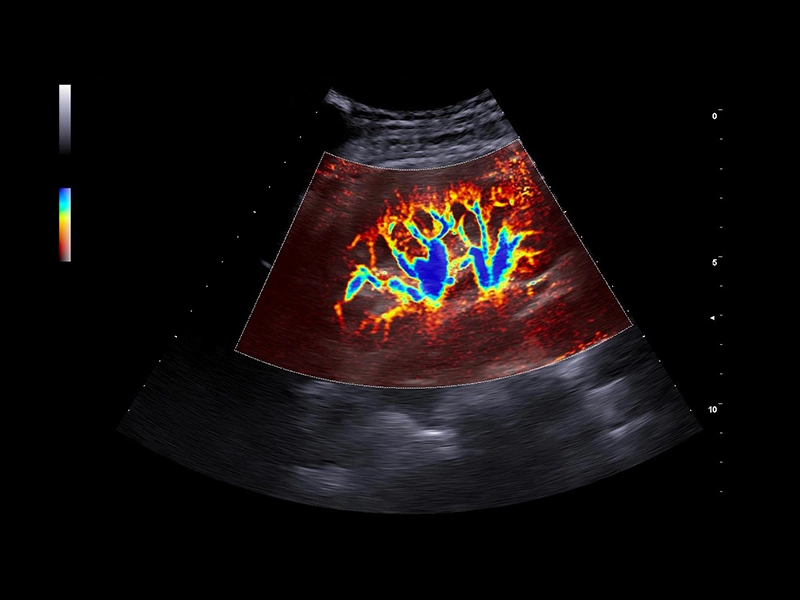
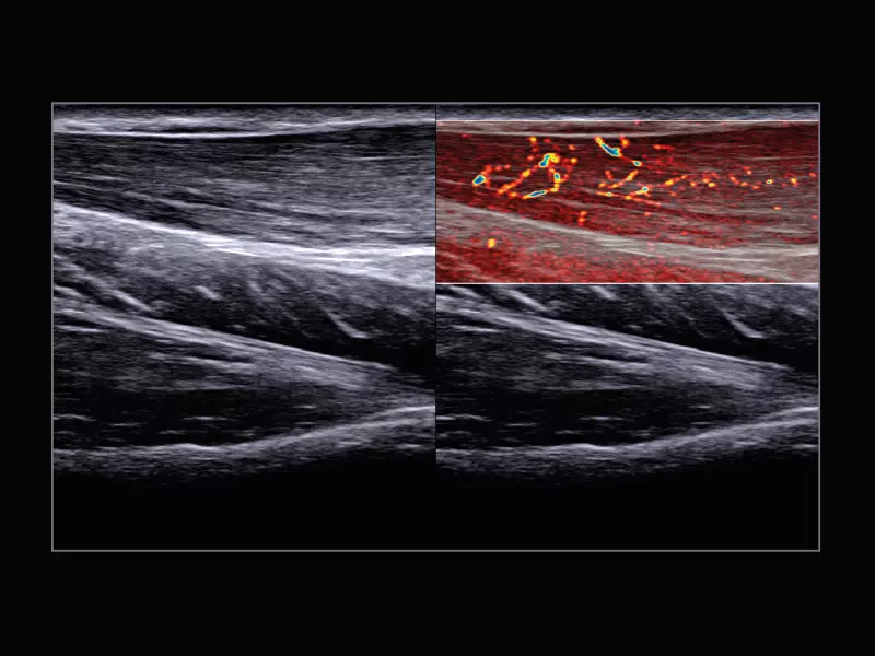
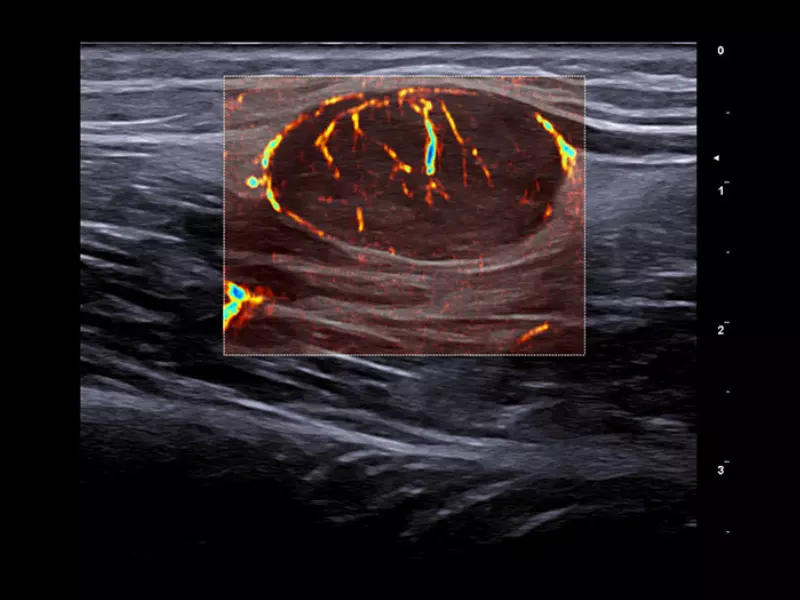
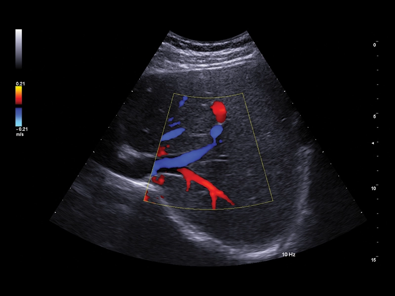
MyLab™E85 GTS Edition - Kidney with microV technology
MyLab™E85 GTS Edition - Kidney with microV technology
MyLab™E85 GTS Edition - Liver with microV technology
MyLab™E85 GTS Edition - Liver with microV technology
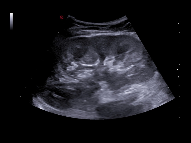
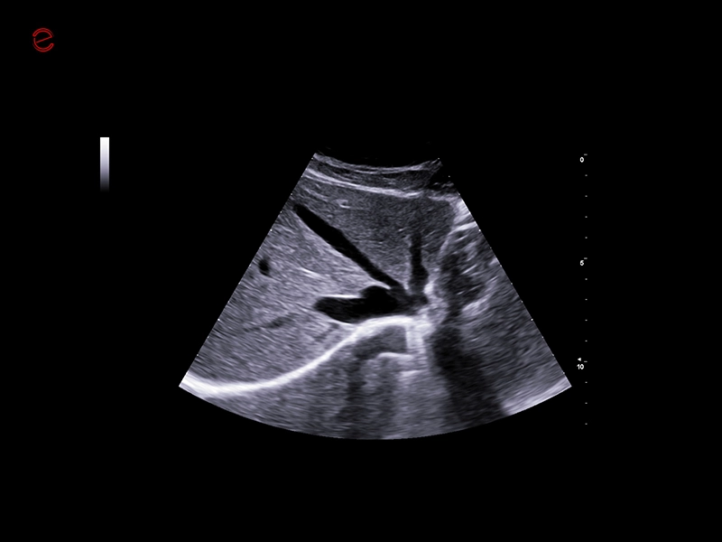
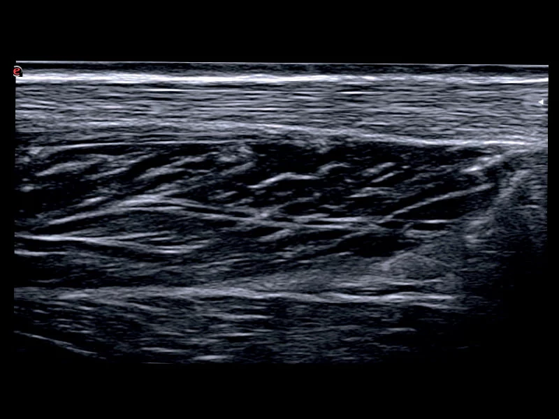
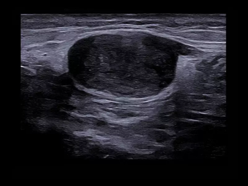
MyLab™E85 GTS Edition - B-Mode image of the kidney
MyLab™E85 GTS Edition - B-Mode image of the kidney
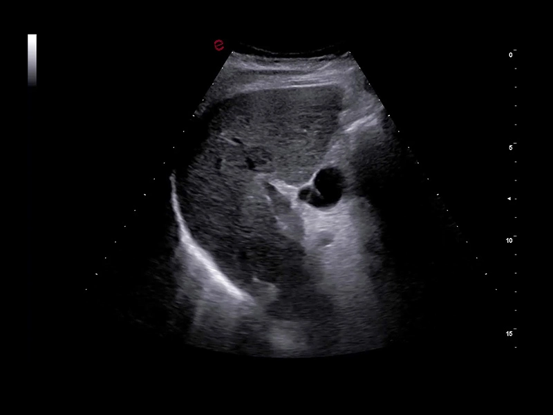
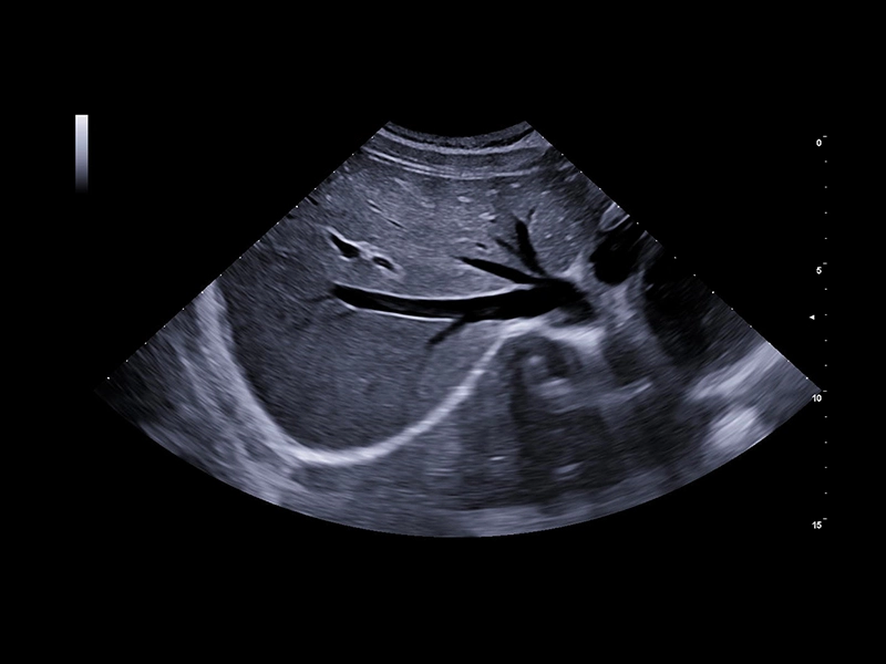
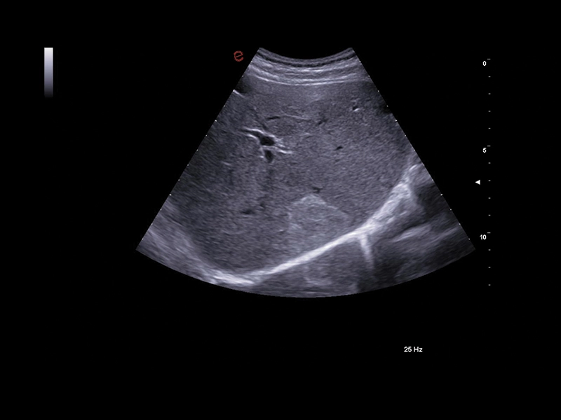
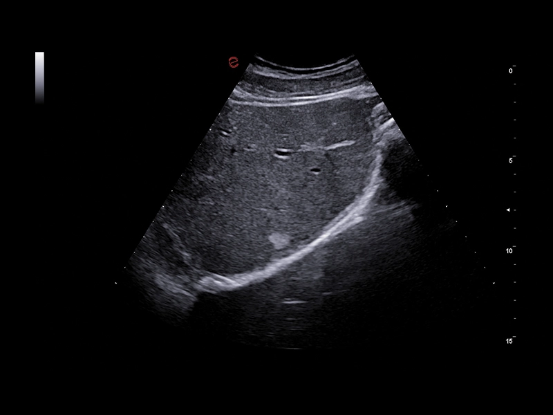
MyLab™E85 GTS Edition - B-Mode image of the liver
MyLab™E85 GTS Edition - B-Mode image of the liver
MyLab™E85 - eDetect technology
MyLab™E85 - eDetect technology
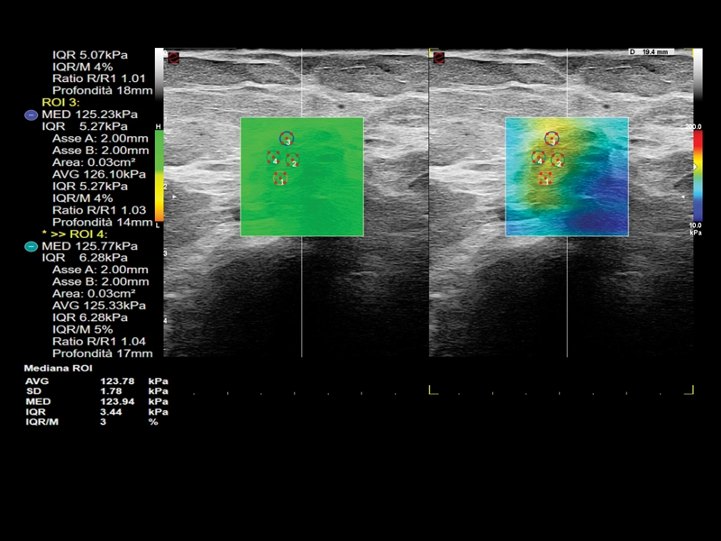
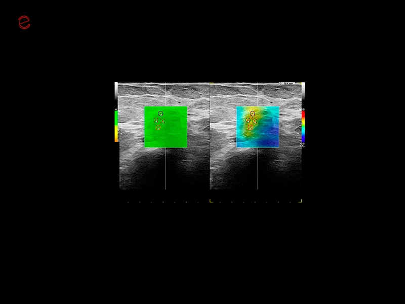
MyLab™E85 - ShearWave Elastography technology
MyLab™E85 - ShearWave Elastography technology
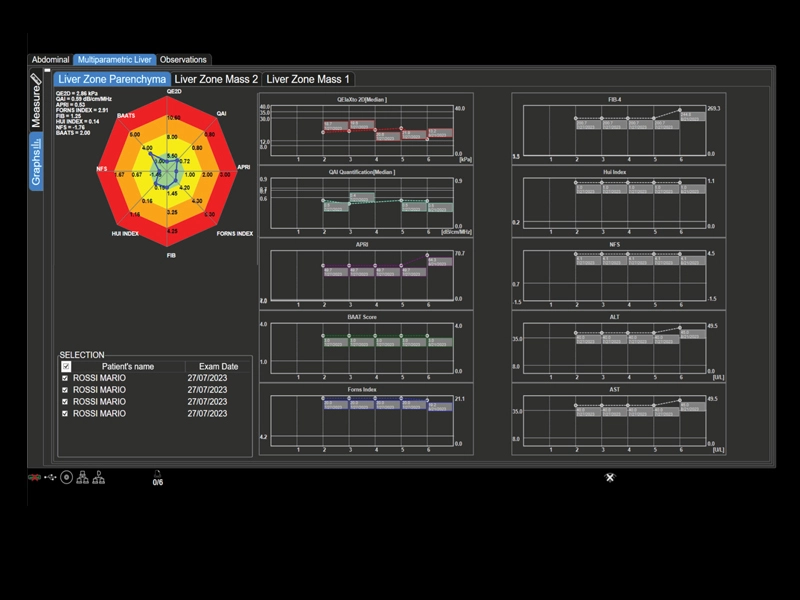
MyLab™E85 - Multiparametric Report
MyLab™E85 - Multiparametric Report
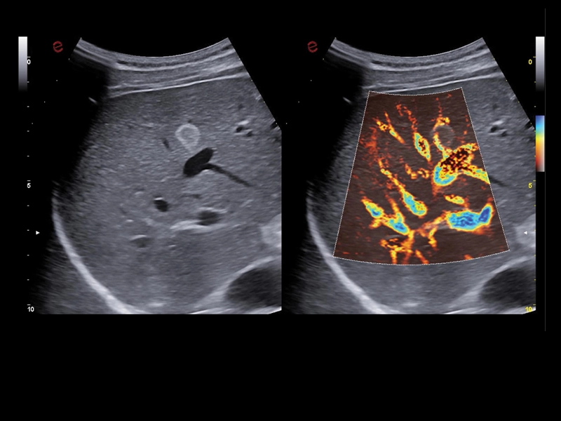
MyLab™E85 - Liver with microV technology
MyLab™E85 - Liver with microV technology
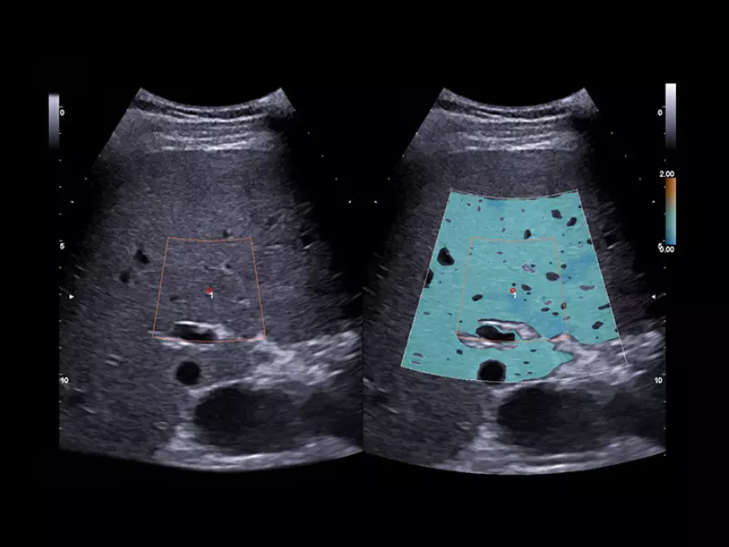
MyLab™E85 - Liver with QSI technology
MyLab™E85 - Liver with QSI technology
MyLab™E85 - Liver with QElaXto 2D technology
MyLab™E85 - Liver with QElaXto 2D technology
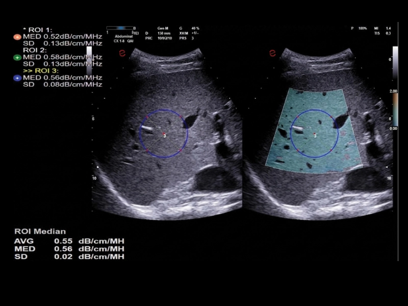
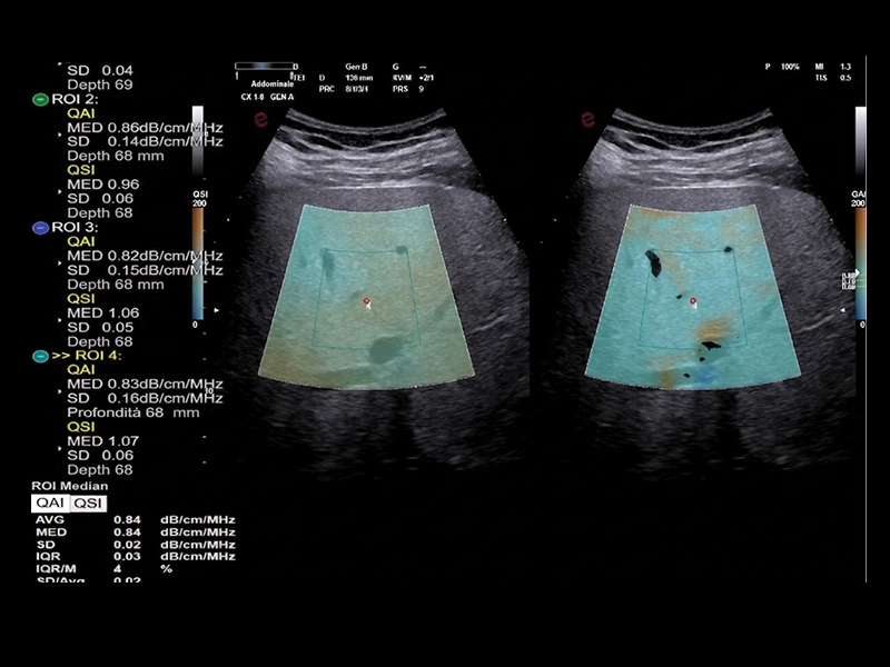
MyLab™E85 - Liver with QAI technology
MyLab™E85 - Liver with QAI technology
MyLab™E85 - Liver CnTI™ Clear with convex probe
MyLab™E85 - Liver CnTI™ Clear with convex probe
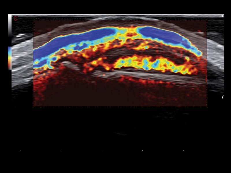
MyLab™E85 - Finger with microV technology
MyLab™E85 - Finger with microV technology
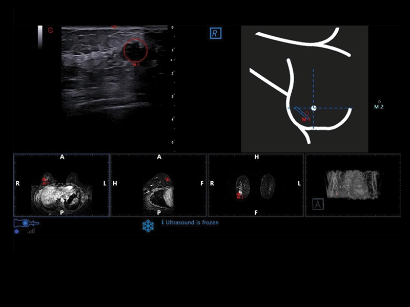
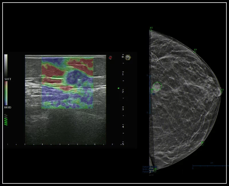
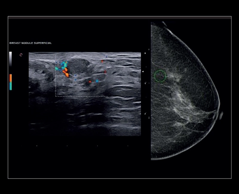
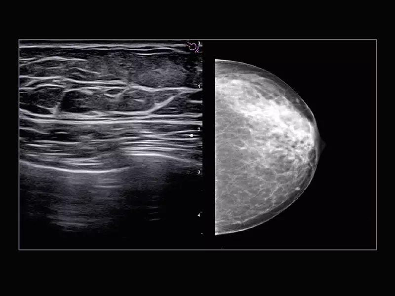
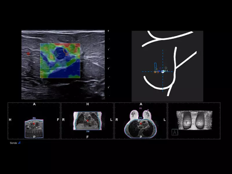
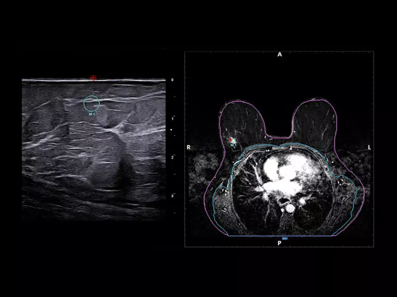
MyLab™E85 - BreastNav™MRI technology
MyLab™E85 - BreastNav™MRI technology
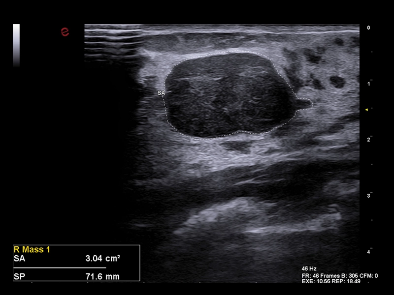
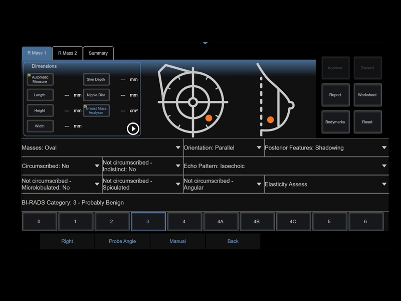
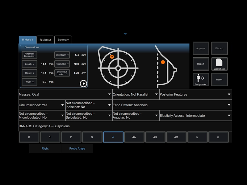
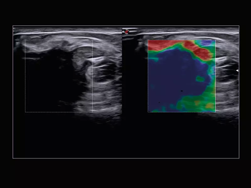
MyLab™E85 - Breast Mass Analyzer (BMA) technology
MyLab™E85 - Breast Mass Analyzer (BMA) technology
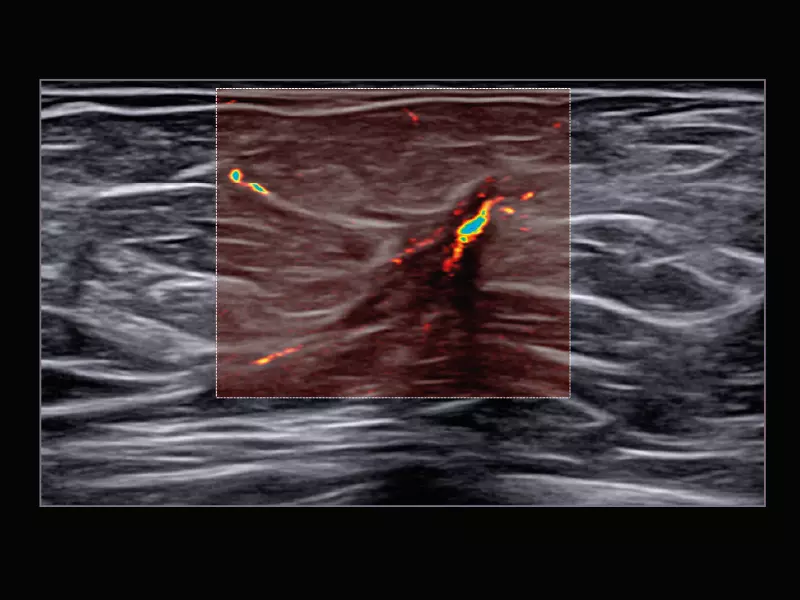
MyLab™E85 - Breast Interactive Workflow (BIW) technology
MyLab™E85 - Breast Interactive Workflow (BIW) technology
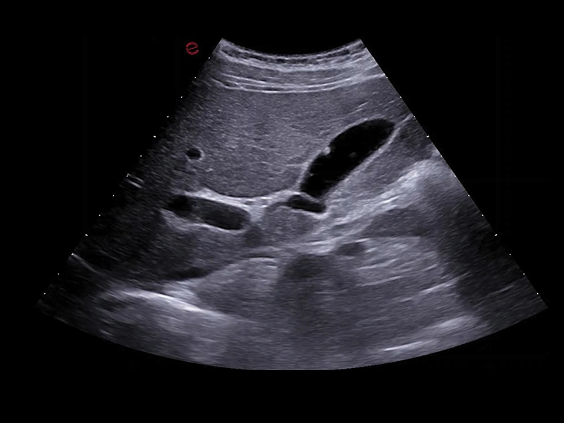
MyLab™E85 - B-Mode of the liver
MyLab™E85 - B-Mode of the liver
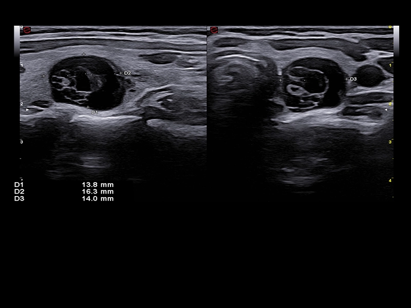
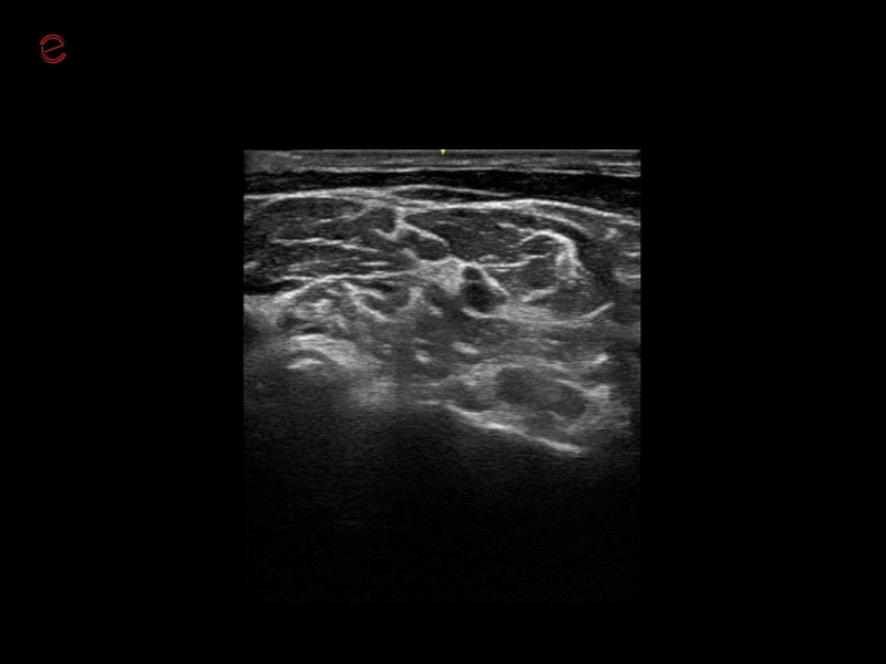
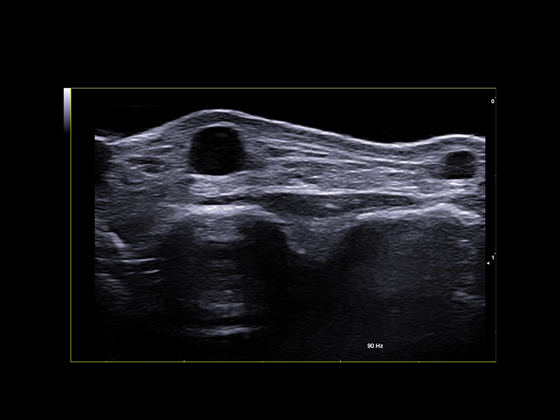
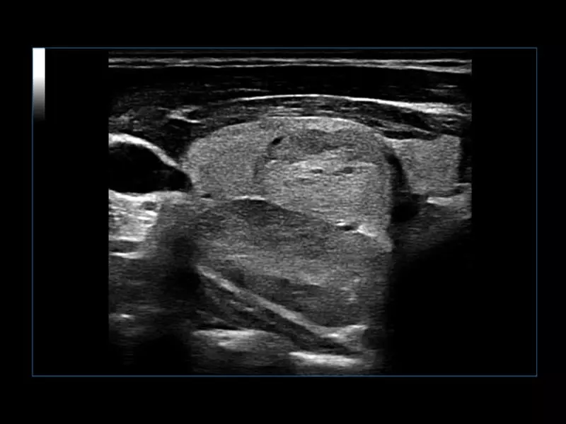
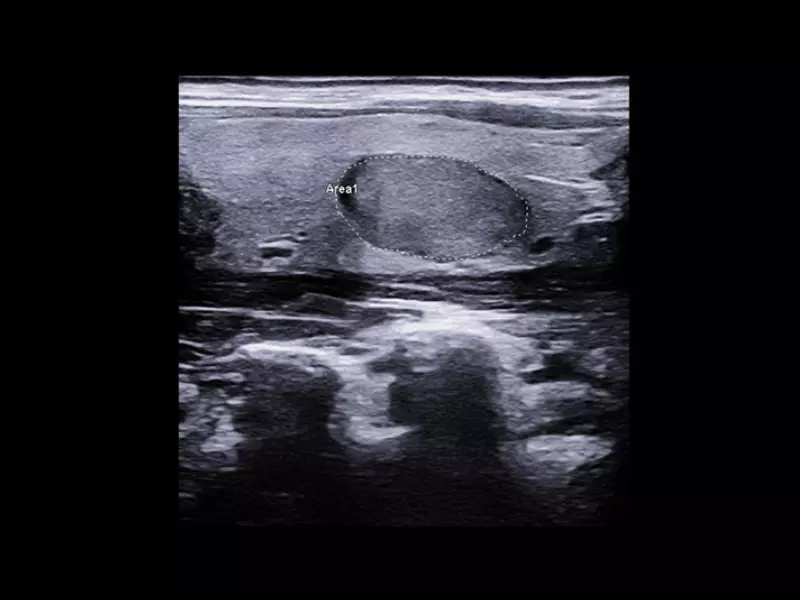
MyLab™E85 - B-Mode image of the thyroid with LMX 3-16
MyLab™E85 - B-Mode image of the thyroid with LMX 3-16
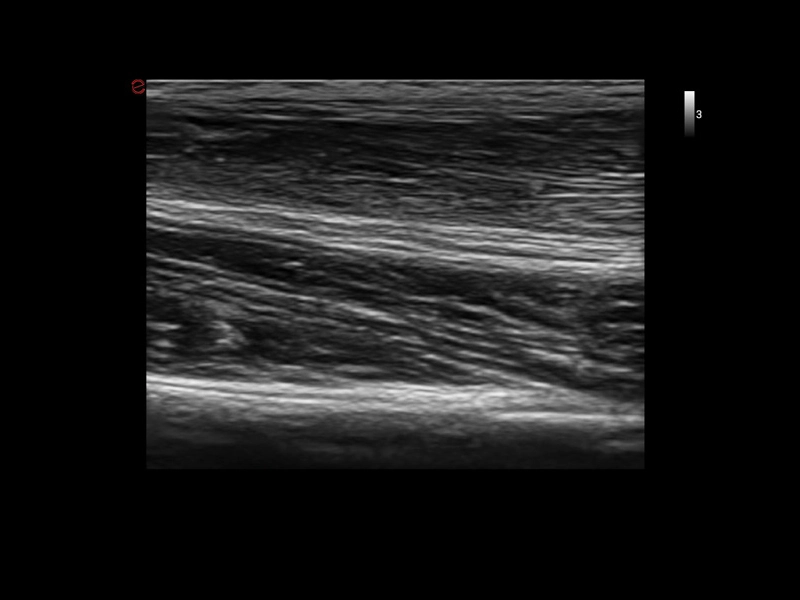
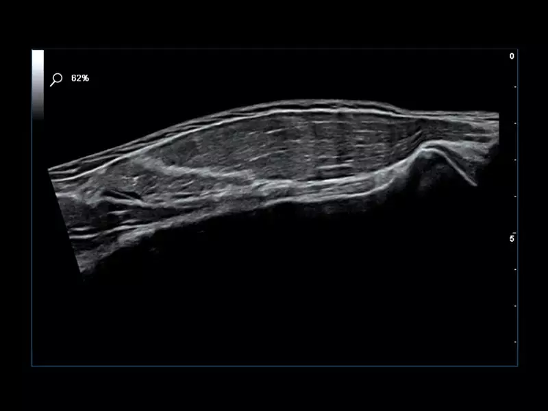
MyLab™E85 - B-Mode image of the quad muscle with LMX 3-16
MyLab™E85 - B-Mode image of the quad muscle with LMX 3-16
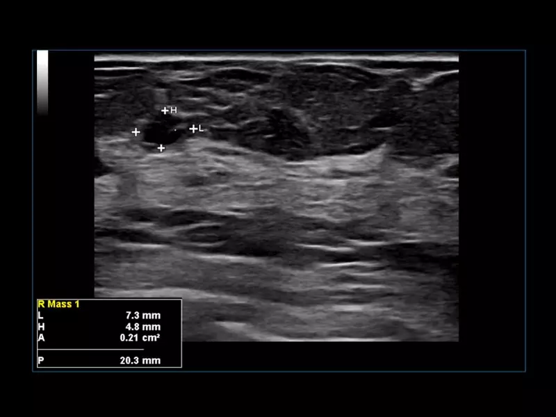
MyLab™E85 - B-Mode image of the breast with LMX 3-16
MyLab™E85 - B-Mode image of the breast with LMX 3-16
MyLab™C30 - Interventional 11 M
MyLab™C30 - Interventional 11 M
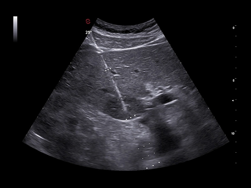
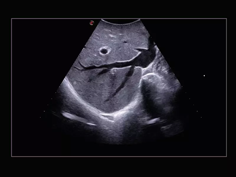
MyLab™C30 - Interventional Liver
MyLab™C30 - Interventional Liver
MyLab™C30 - Interventional 09 M
MyLab™C30 - Interventional 09 M
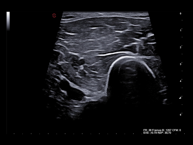
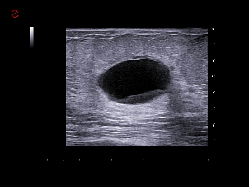
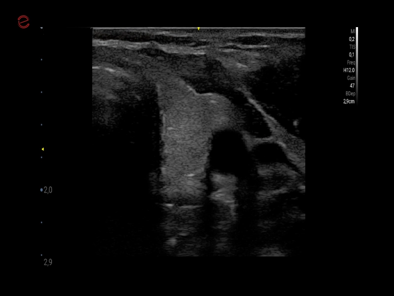
MyLab™C30 - Interventional B-Mode - Testis
MyLab™C30 - Interventional B-Mode - Testis
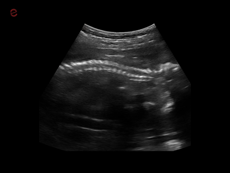
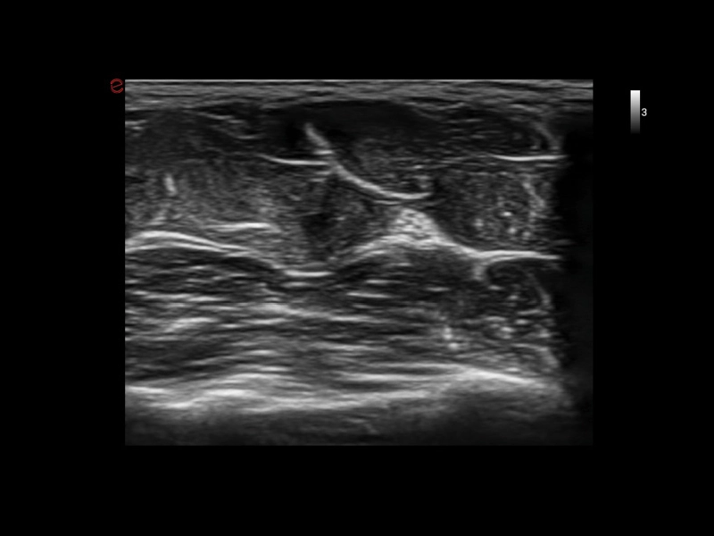
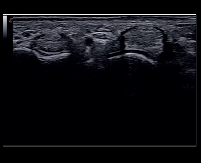
MyLab™C30 - Interventional Vertebral
MyLab™C30 - Interventional Vertebral
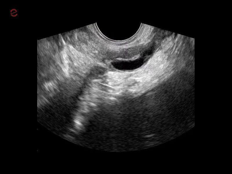
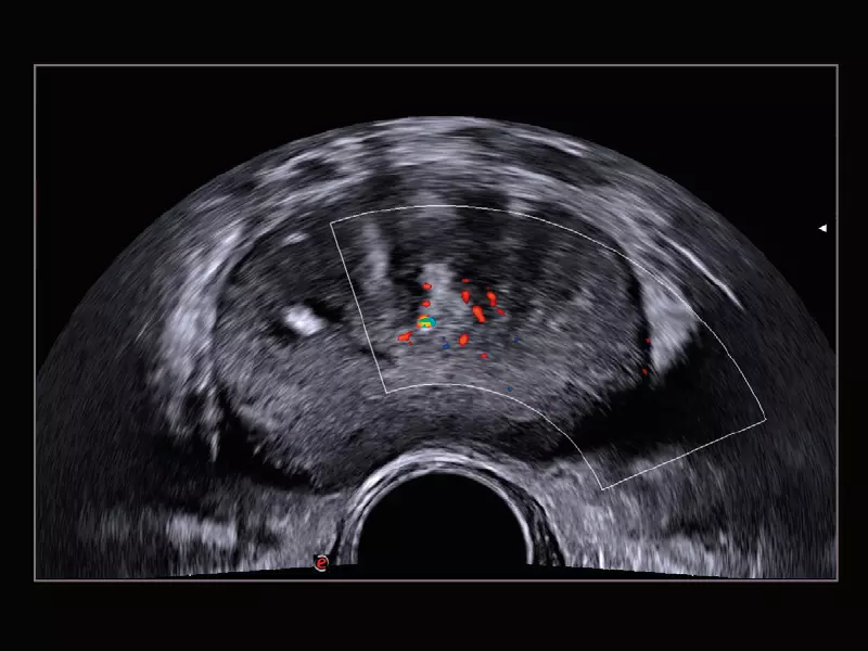
MyLab™C30 - Interventional Prostate
MyLab™C30 - Interventional Prostate
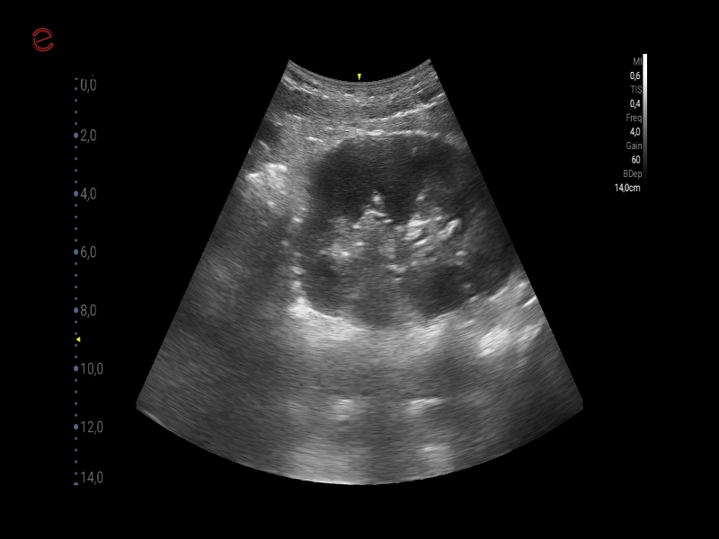
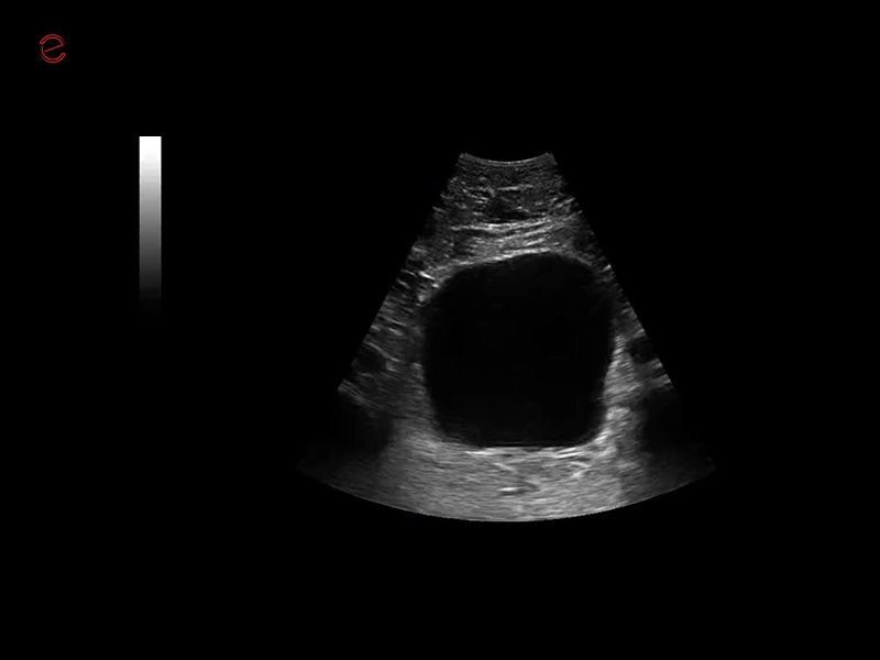
MyLab™C30 - Interventional B-Mode - Kidney
MyLab™C30 - Interventional B-Mode - Kidney
MyLab™C30 - Interventional 03 M
MyLab™C30 - Interventional 03 M
MyLab™C30 - Interventional 02 M
MyLab™C30 - Interventional 02 M
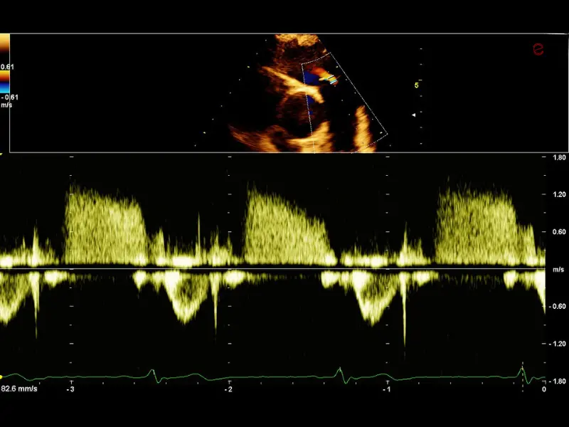
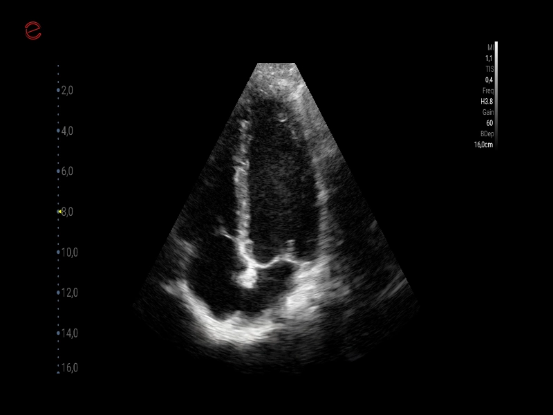
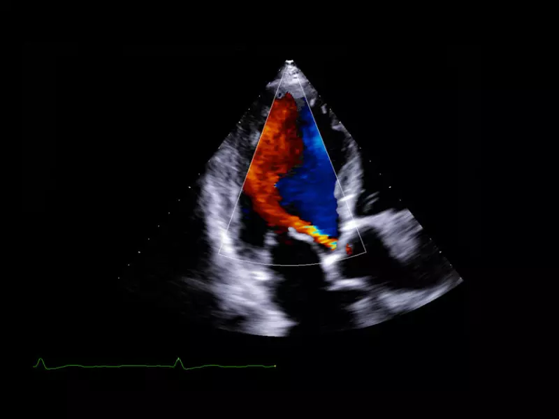
MyLab™C30 - TVM - 4CH
MyLab™C30 - TVM - 4CH
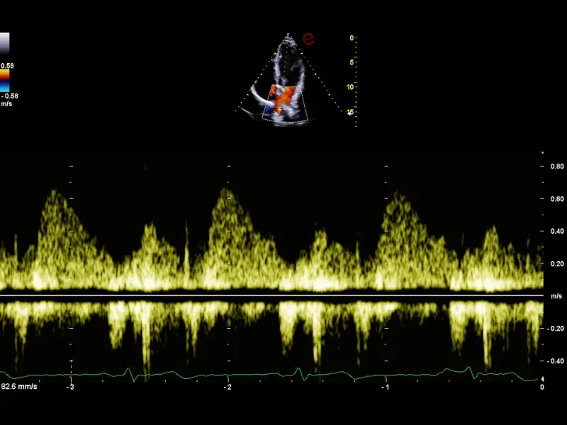
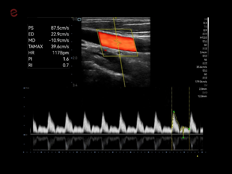
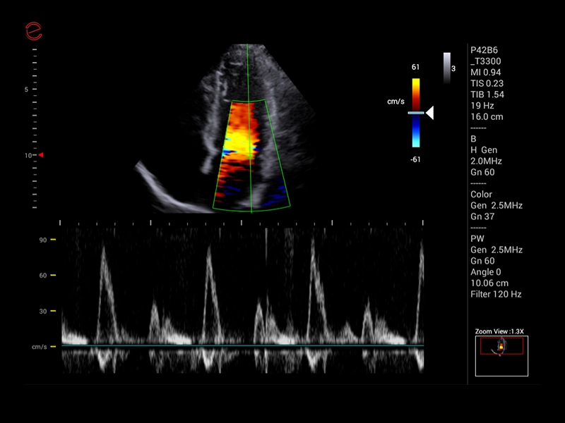
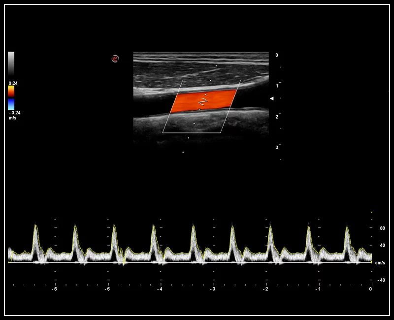
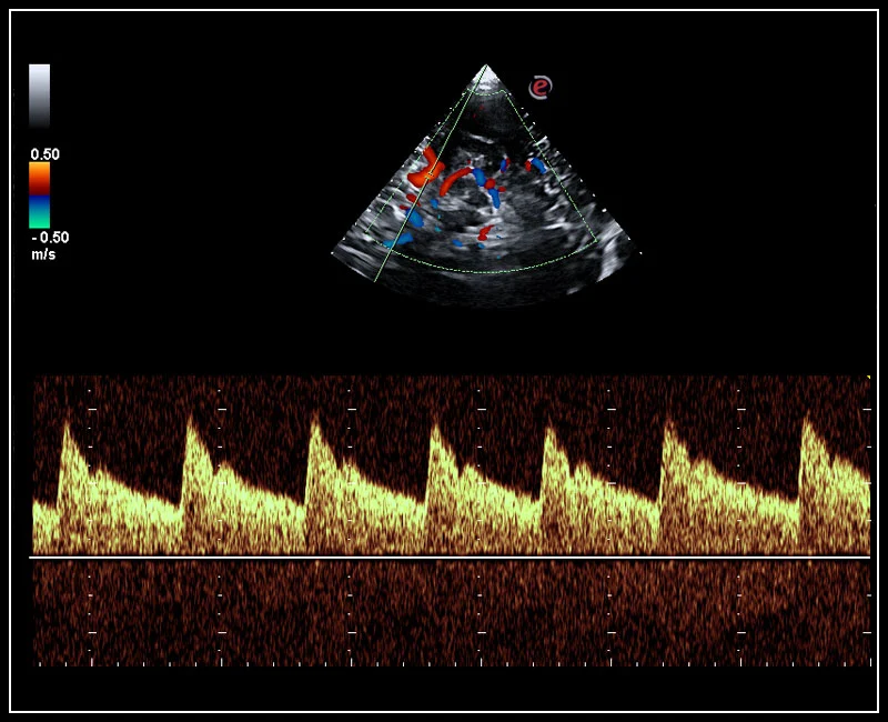
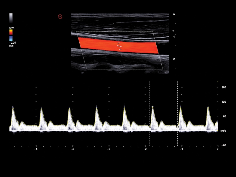
MyLab™C30 - Polm - Doppler
MyLab™C30 - Polm - Doppler
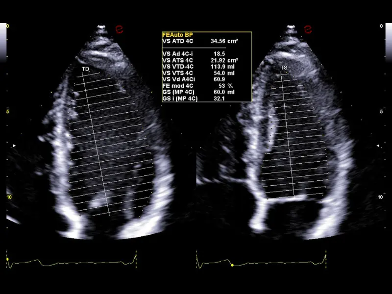
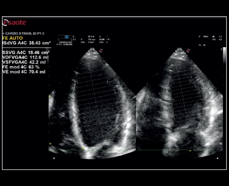
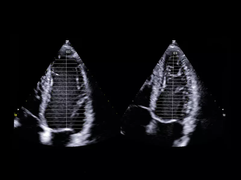
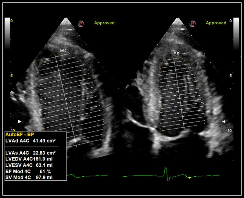
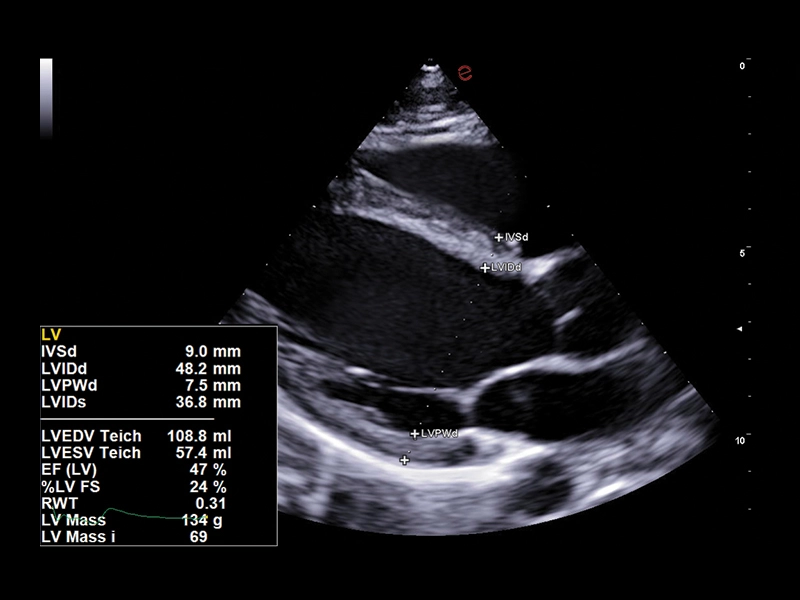
MyLab™C30 - FE
MyLab™C30 - FE
MyLab™C30 - Doppler - CW
MyLab™C30 - Doppler - CW
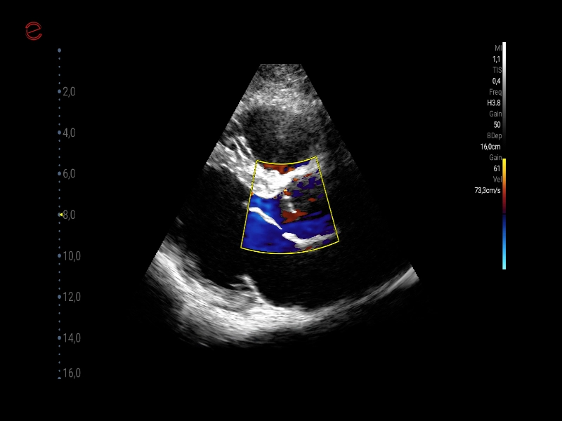
MyLab™C30 - CFM - TR
MyLab™C30 - CFM - TR
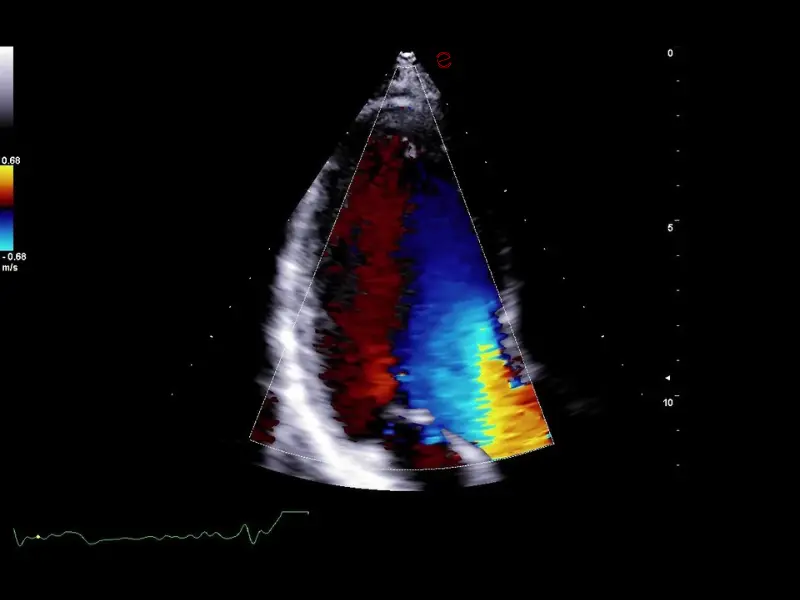
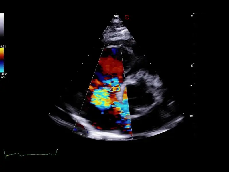
MyLab™C30 - CFM - 3CH
MyLab™C30 - CFM - 3CH
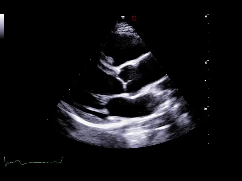
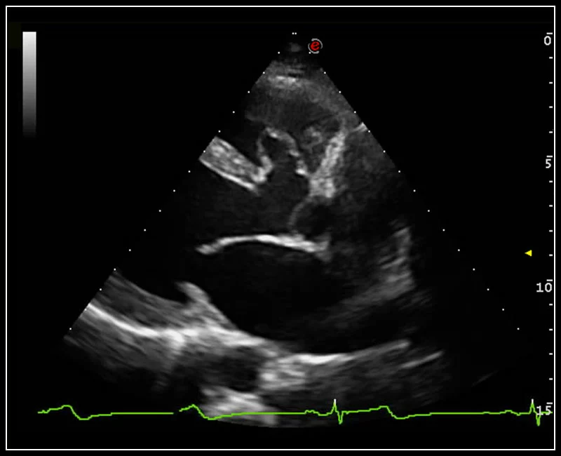
MyLab™C30 - Bmode - Plax
MyLab™C30 - Bmode - Plax
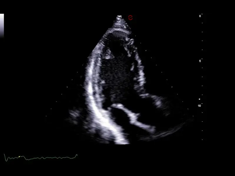
MyLab™C30 - Bmode - 3CH
MyLab™C30 - Bmode - 3CH
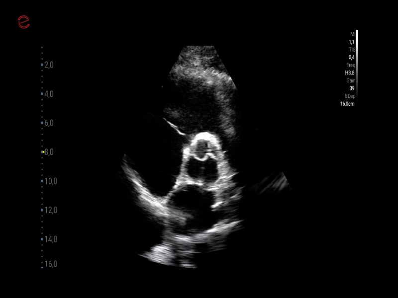
MyLab™C30 - Aorta
MyLab™C30 - Aorta
MyLab™C30 - QElaxto 2D liver
MyLab™C30 - QElaxto 2D liver
MyLab™C30 - MICROV-NEW
MyLab™C30 - MICROV-NEW
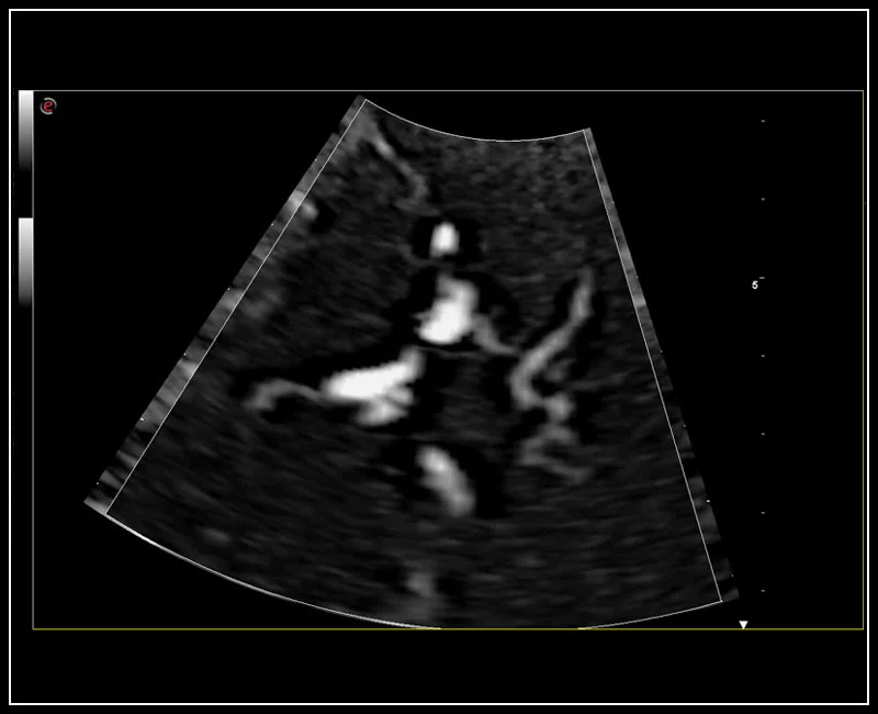
MyLab™C30 - MCA
MyLab™C30 - MCA
MyLab™C30 - 4D VC2
MyLab™C30 - 4D VC2
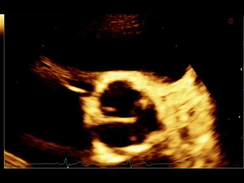
MyLab™C30 - Hyper
MyLab™C30 - Hyper
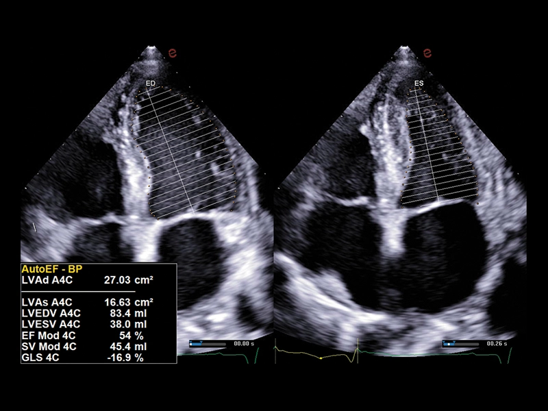
MyLab™C30 - 4d strain
MyLab™C30 - 4d strain
MyLab™C30 - General Imaging
MyLab™C30 - General Imaging
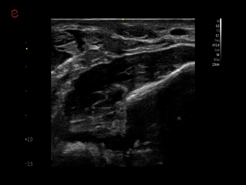
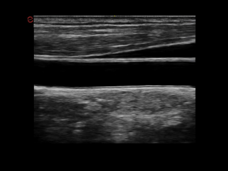
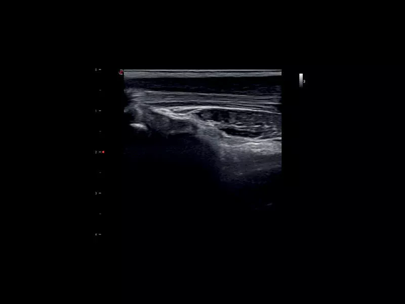
MyLab™C30 - MSK nerve
MyLab™C30 - MSK nerve
MyLab™C30 - Liver QAI
MyLab™C30 - Liver QAI
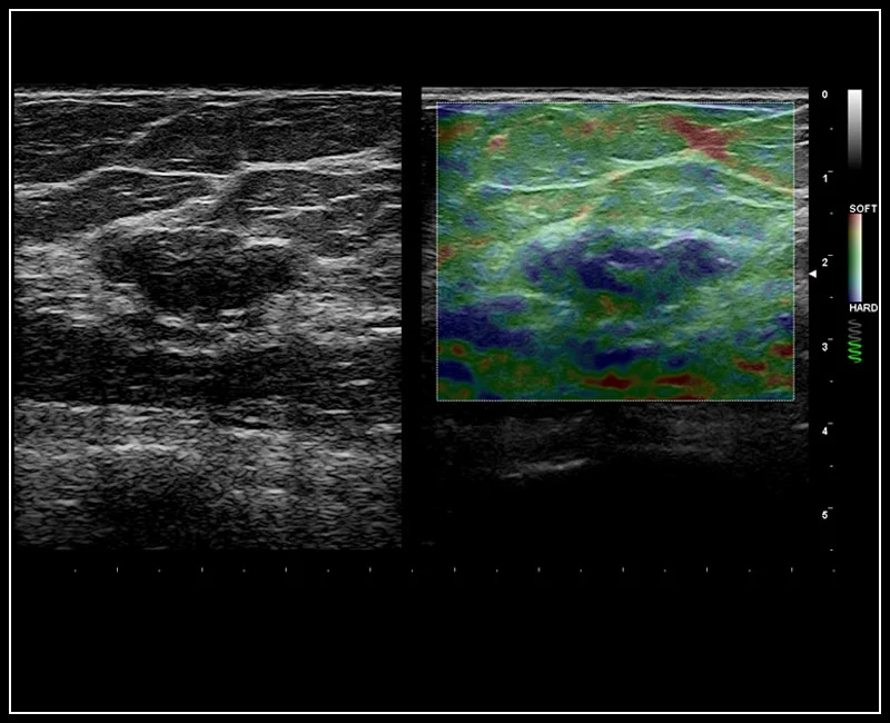
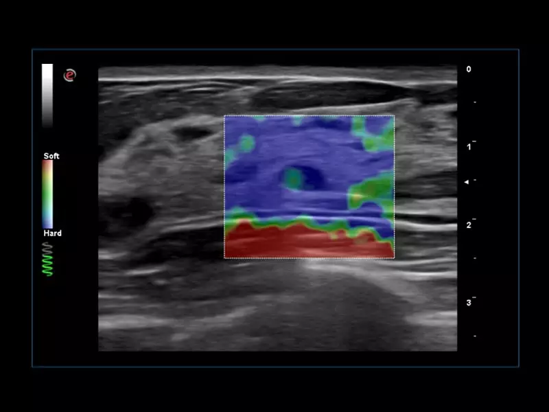
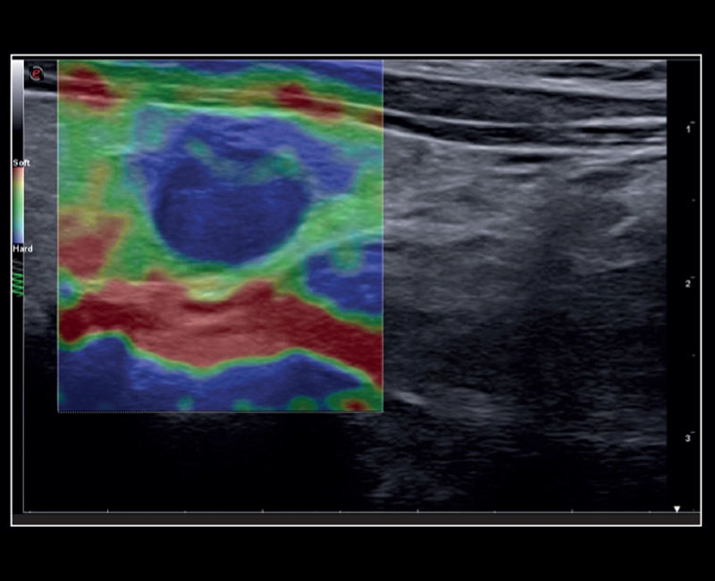
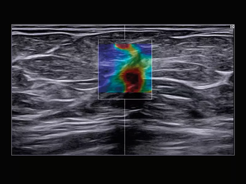
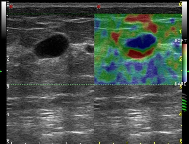
MyLab™C30 - SWE breast
MyLab™C30 - SWE breast
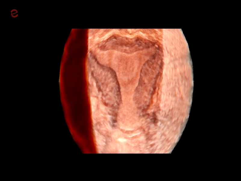
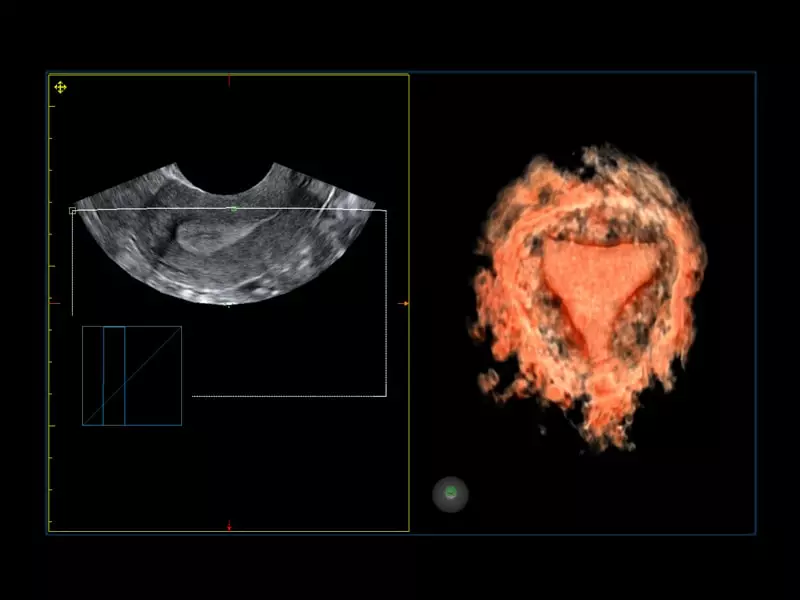
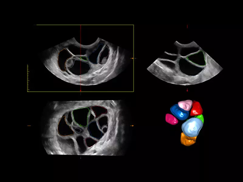
MyLab™C30 - Gyn 3D
MyLab™C30 - Gyn 3D
MyLab™C30 - General Imaging
MyLab™C30 - General Imaging
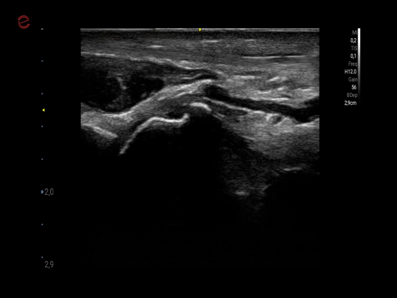
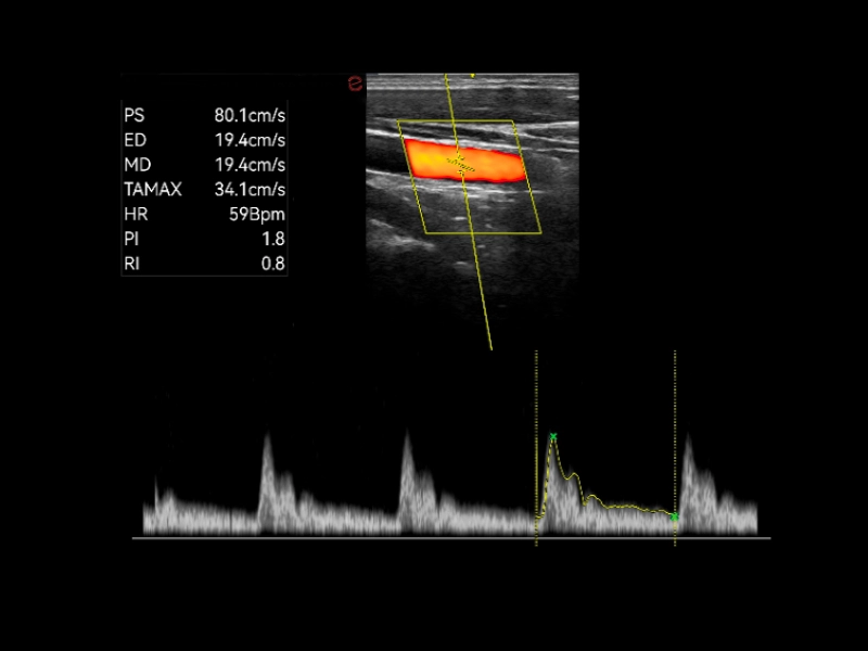
MyLab™C30 - Vertebral PW
MyLab™C30 - Vertebral PW
MyLab™C30 - Prostate median plan axial view TLC 3-13
MyLab™C30 - Prostate median plan axial view TLC 3-13
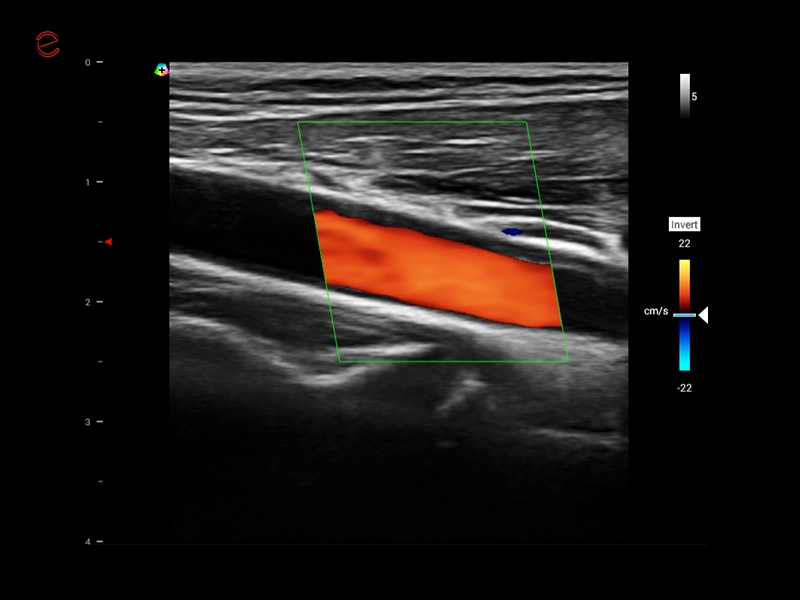
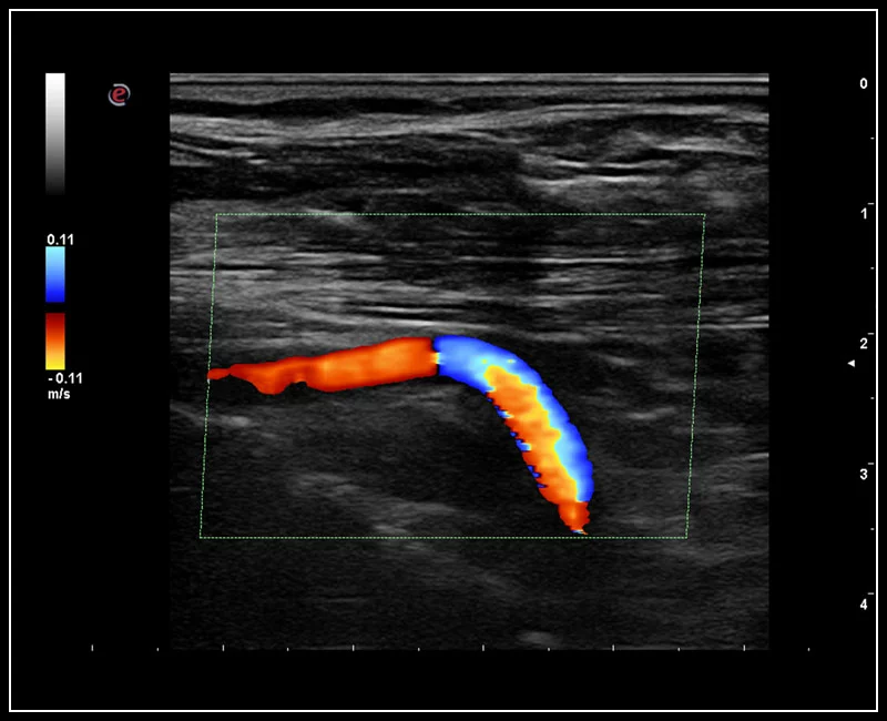
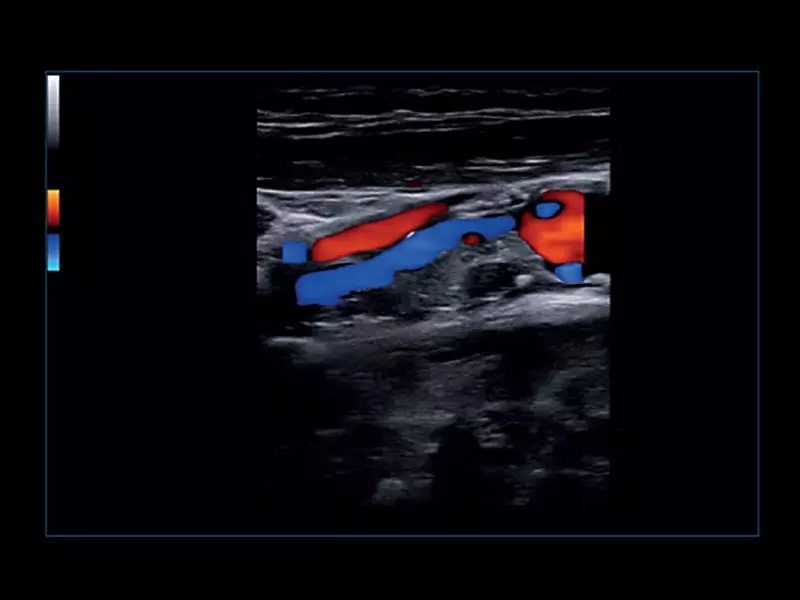
MyLab™C25 - TSA-CFM
MyLab™C25 - TSA-CFM
MyLab™C25 - Bmode
MyLab™C25 - Bmode
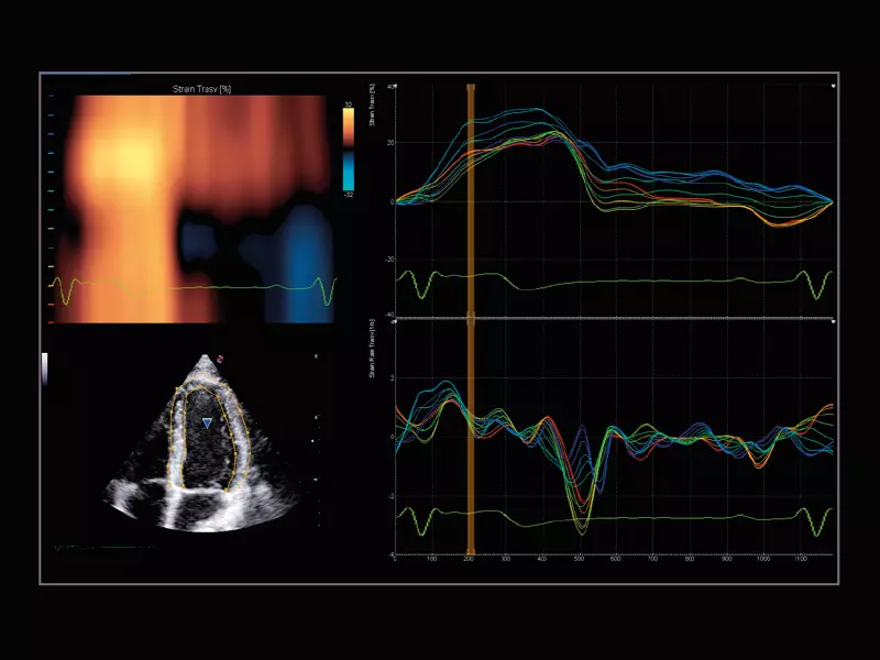
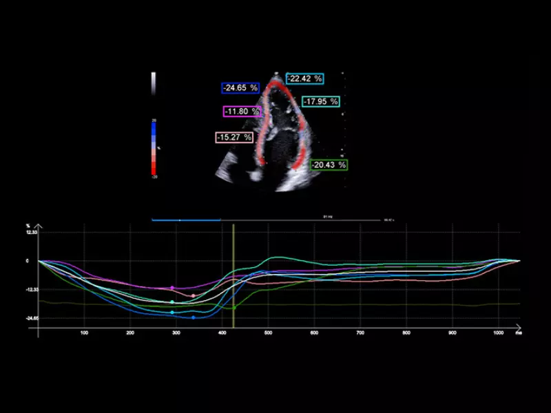
MyLab™C25 - Xstrain
MyLab™C25 - Xstrain
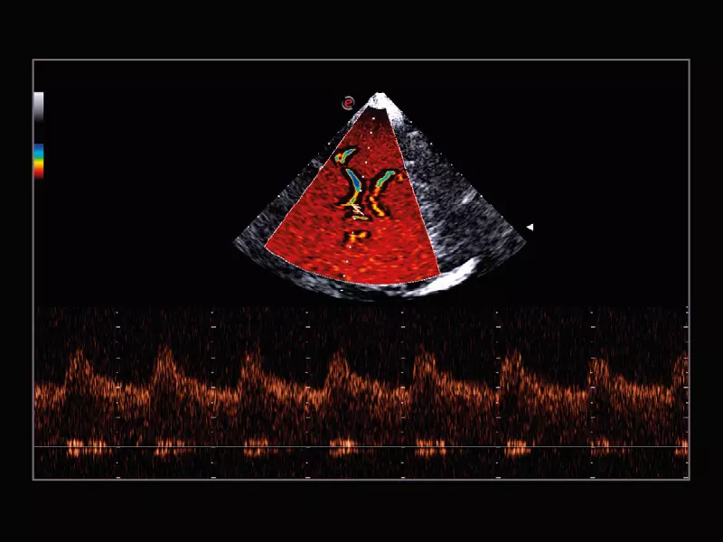
MyLab™C25 - TP-IQ
MyLab™C25 - TP-IQ
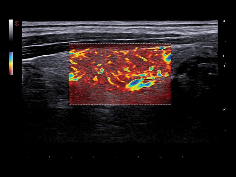
MyLab™C25 - MICRO-V-thyroid-linear
MyLab™C25 - MICRO-V-thyroid-linear
MyLab™C25 - IHX muscle
MyLab™C25 - IHX muscle
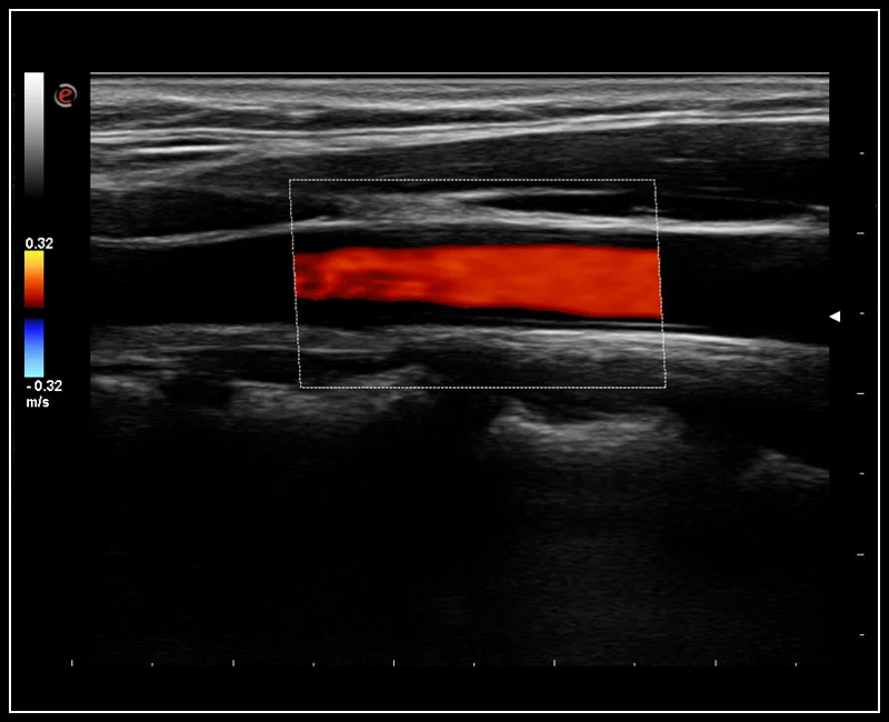
MyLab™C25 - X-FLOW 2
MyLab™C25 - X-FLOW 2
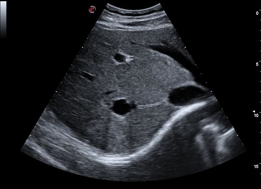
MyLab™C25 - BMODE
MyLab™C25 - BMODE
MyLab™C25 - C25
MyLab™C25 - C25
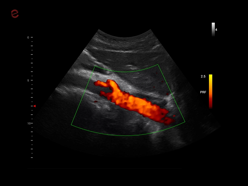
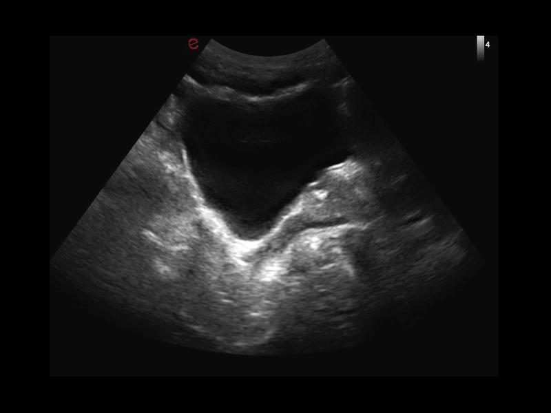
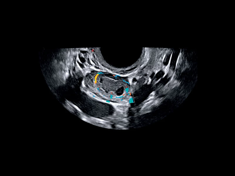
MyLab™C25 - Mioma PD
MyLab™C25 - Mioma PD
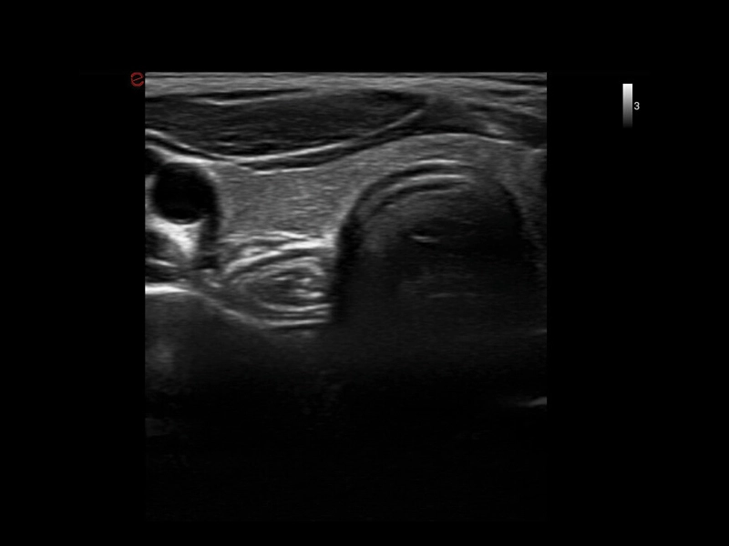
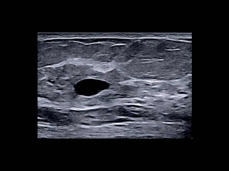
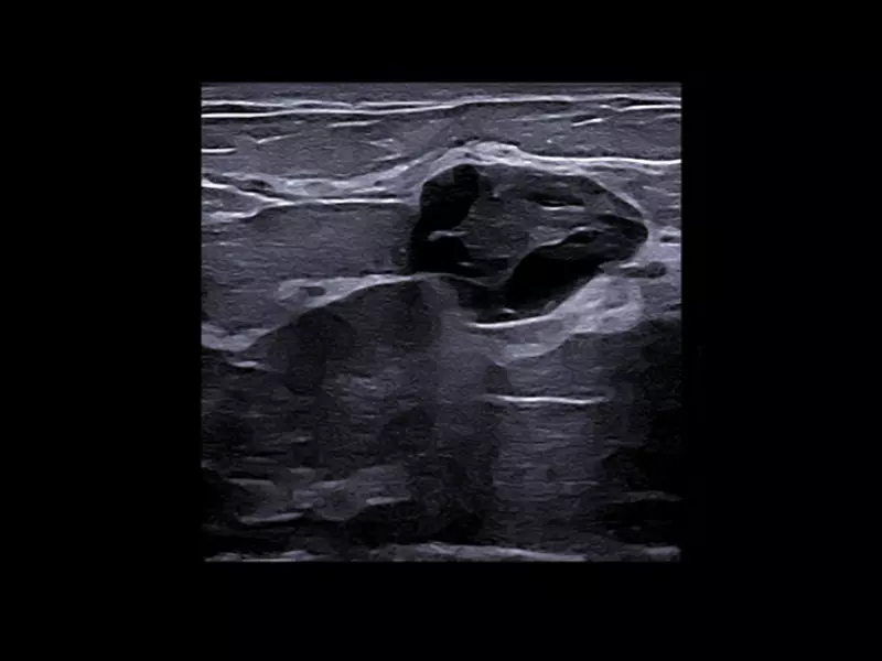
MyLab™C25 - Breast lesion
MyLab™C25 - Breast lesion
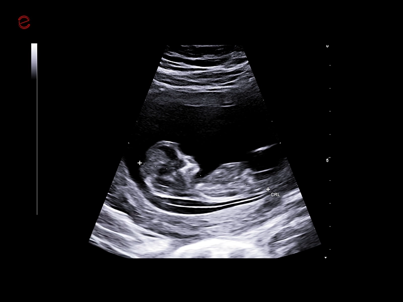
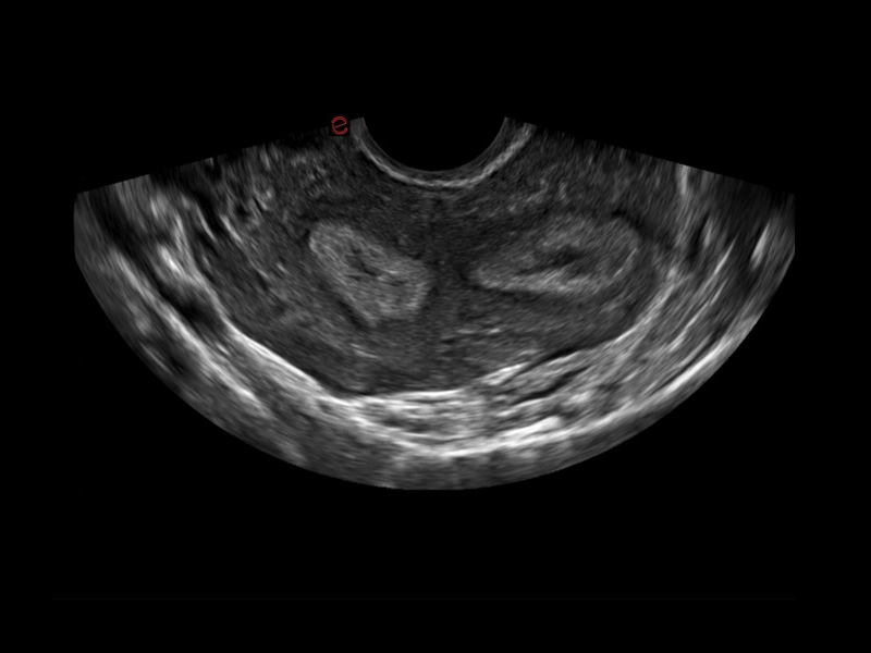
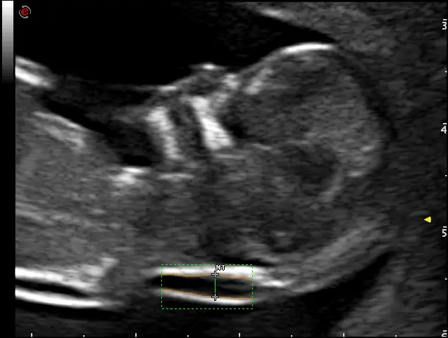
MyLab™C25 - CRL
MyLab™C25 - CRL
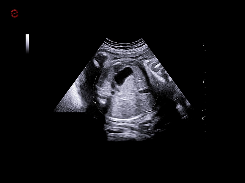
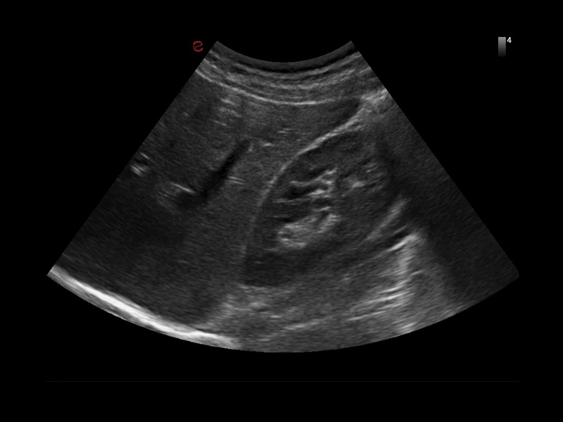
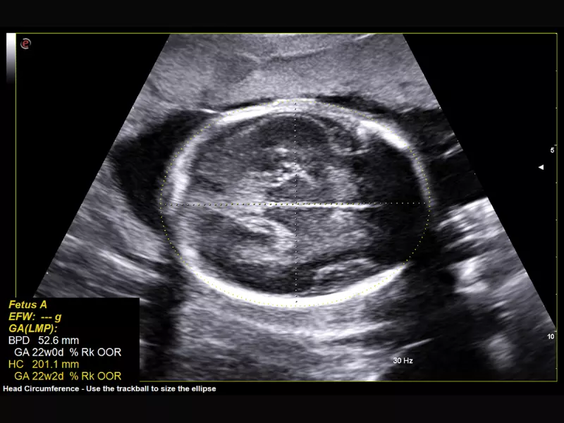
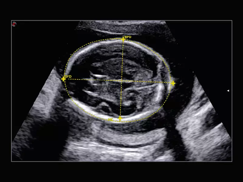
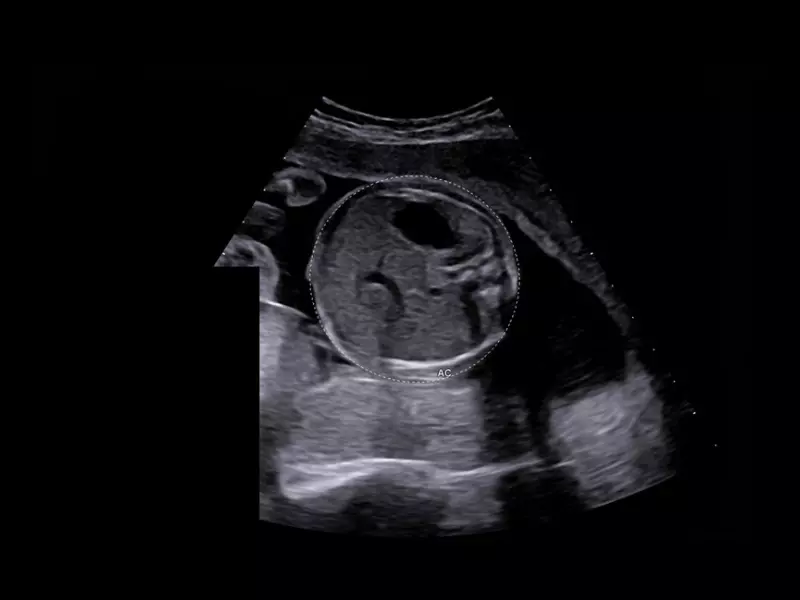
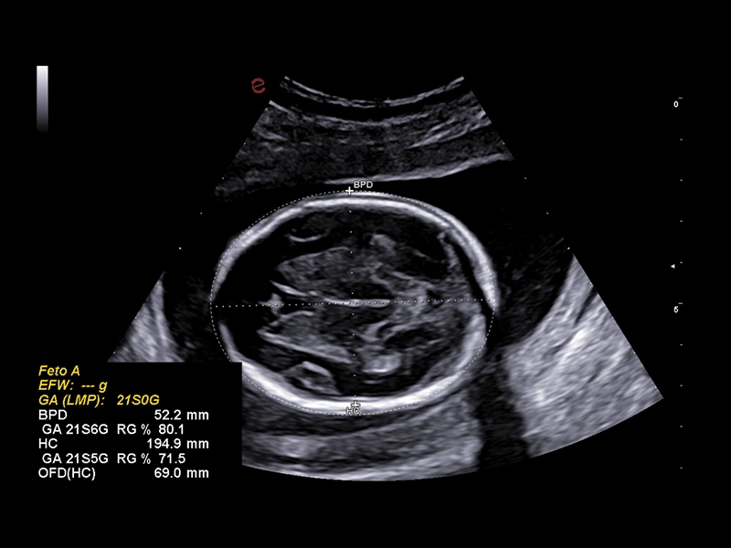
MyLab™C25 - AUTO OB
MyLab™C25 - AUTO OB
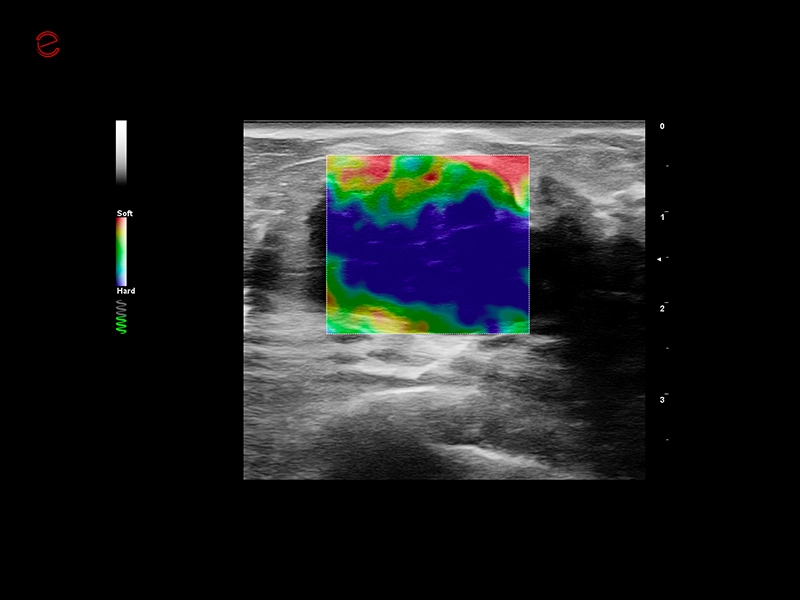
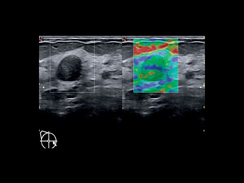
MyLab™C25 - ElaXto in breast
MyLab™C25 - ElaXto in breast
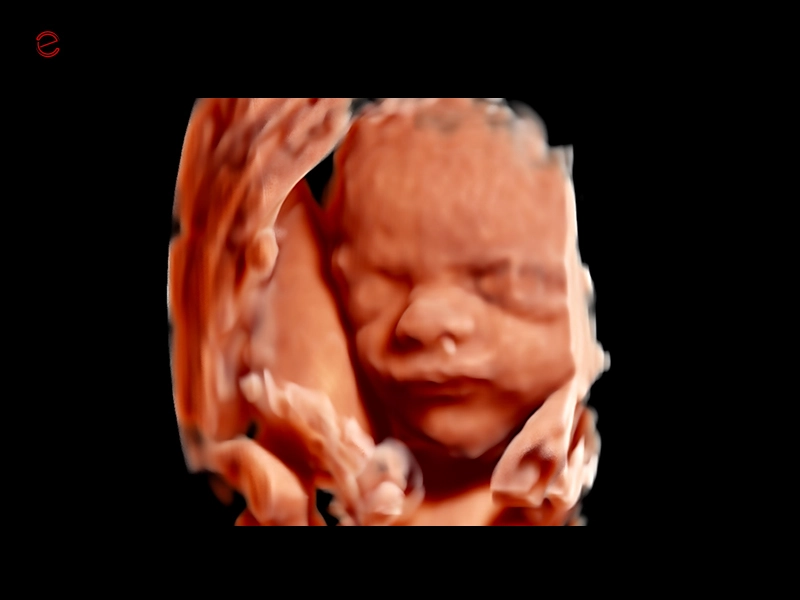
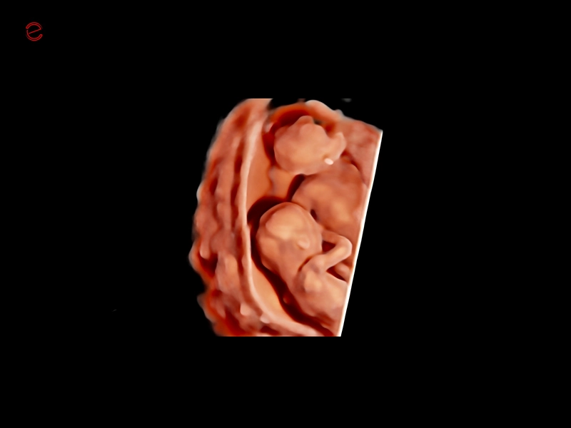
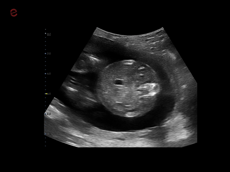
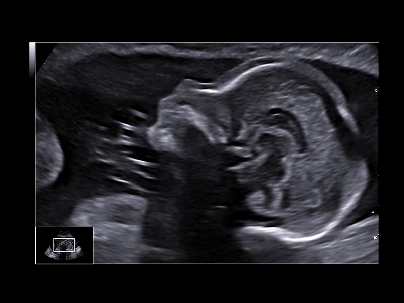
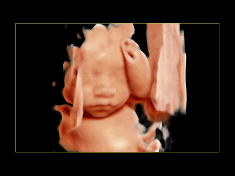
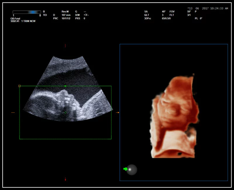
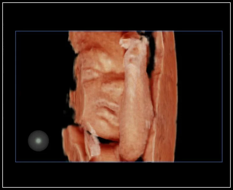
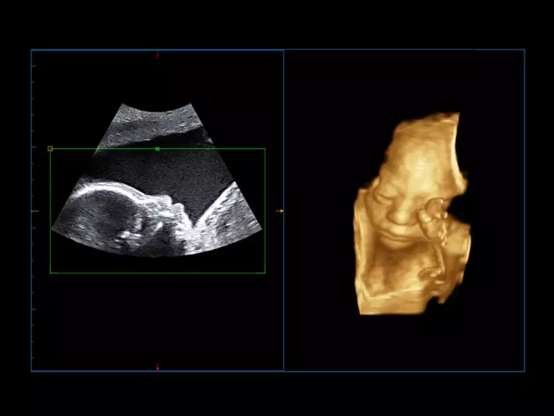
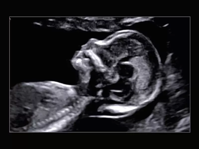
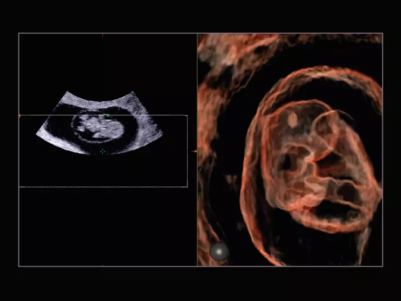
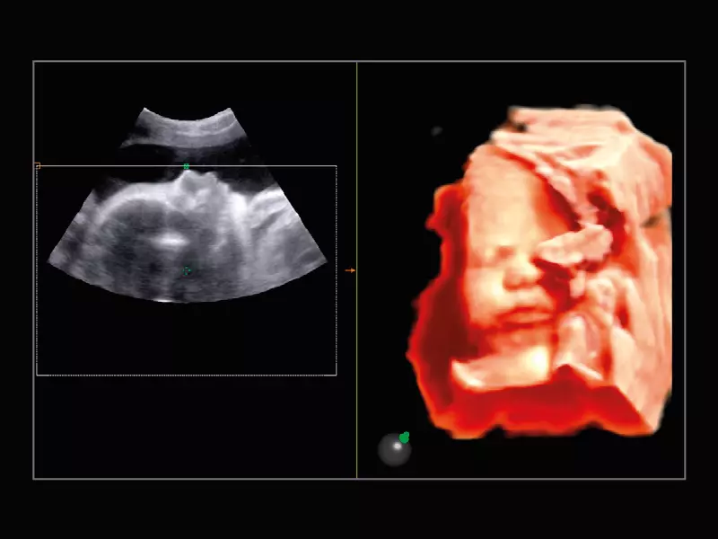
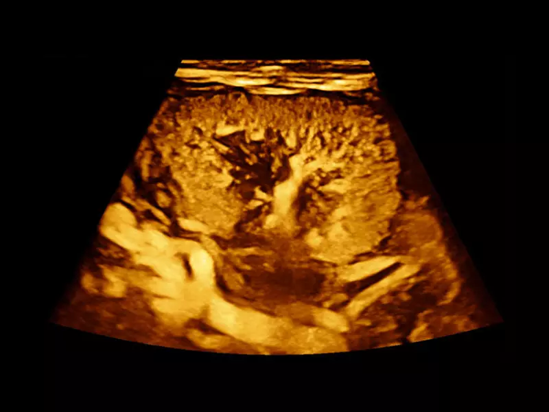
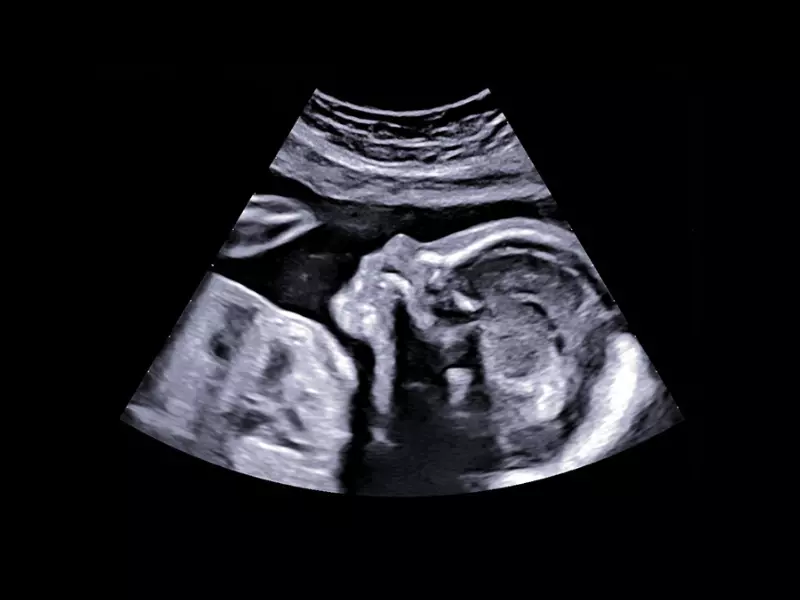
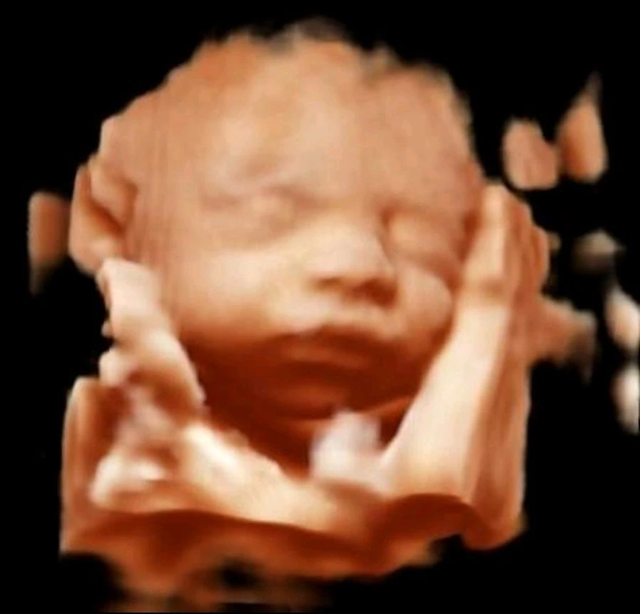
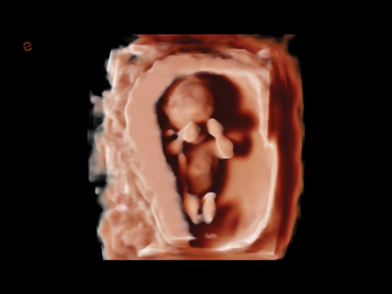
MyLab™C25 - Baby face
MyLab™C25 - Baby face
Q7 - CV Cardio B-mode
Q7 - CV Cardio B-mode
Q7 - CV Cardio B Mode 2
Q7 - CV Cardio B Mode 2
Q7 - CV Cardio
Q7 - CV Cardio
Q7 - CV Vascular
Q7 - CV Vascular
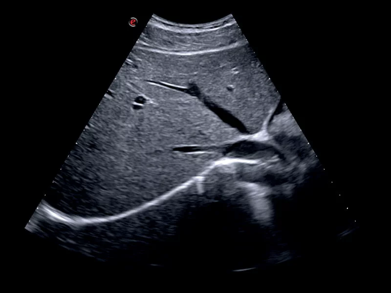
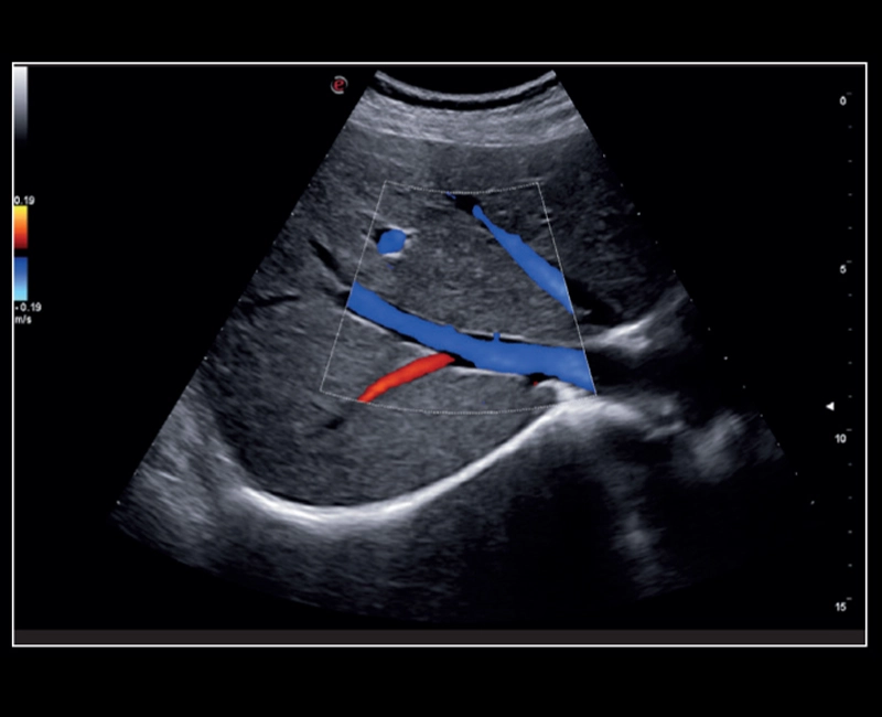
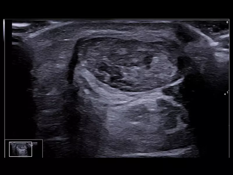
Q7 - GI Kidney
Q7 - GI Kidney
Q7 - GI MSK
Q7 - GI MSK
Q7 - GI Shoulder
Q7 - GI Shoulder
Q7 - GI Small parts
Q7 - GI Small parts
Q7 - POC 2
Q7 - POC 2
Q7 - POC
Q7 - POC
Q7 - WH OB
Q7 - WH OB
Q7 - WH Spine
Q7 - WH Spine
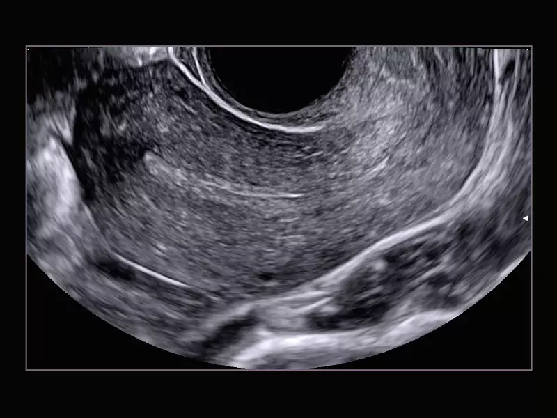
Q7 - WH Uterus
Q7 - WH Uterus
Q7 - Carotid
Q7 - Carotid
Q7 - Doppler
Q7 - Doppler
Q7 - Endo
Q7 - Endo
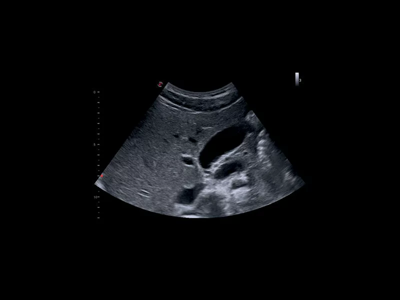
Q7 - Abdominal 02
Q7 - Abdominal 02
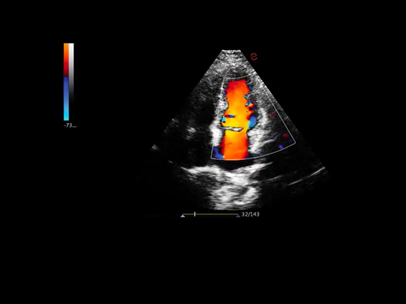
Q7 - Cardio CFM
Q7 - Cardio CFM
Q7 - Abdominal 01
Q7 - Abdominal 01
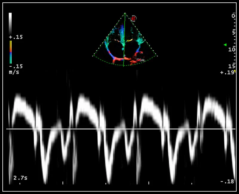
MyLab™X1 Go - CV Doppler 1
MyLab™X1 Go - CV Doppler 1
MyLab™X1 Go - CV Vascular
MyLab™X1 Go - CV Vascular
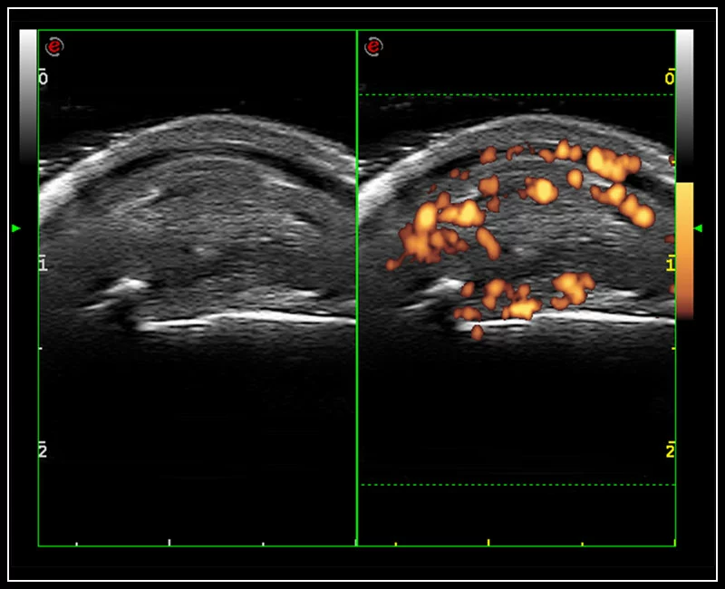
MyLab™X1 Go - GI Abdo CFM
MyLab™X1 Go - GI Abdo CFM
MyLab™X1 Go - GI Kidney
MyLab™X1 Go - GI Kidney
MyLab™X1 Go - GI MSK
MyLab™X1 Go - GI MSK
MyLab™X1 Go - POC 1
MyLab™X1 Go - POC 1
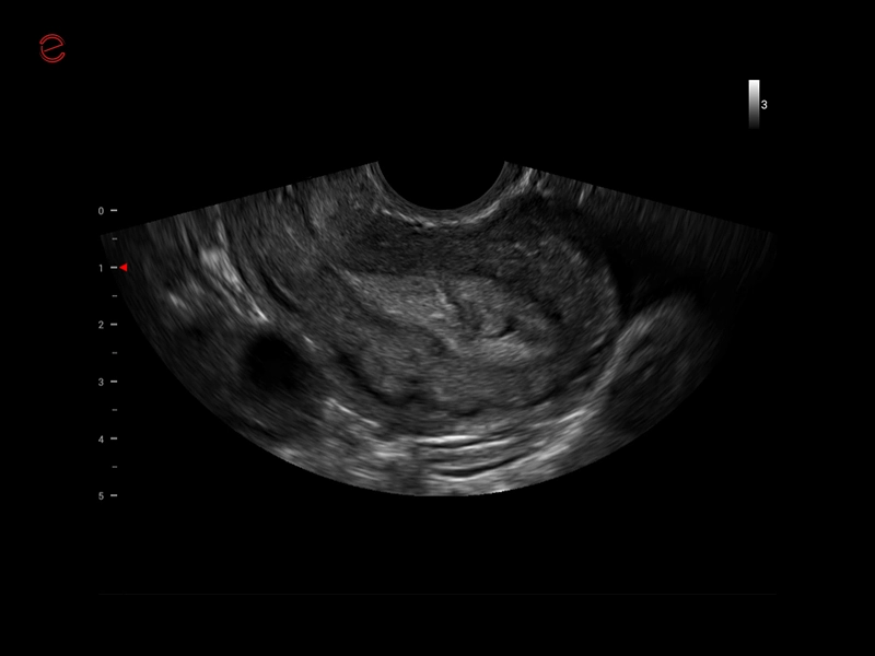
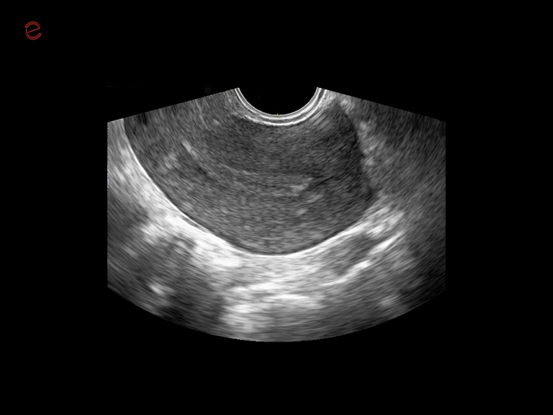
MyLab™X1 Go - WH Endometrium
MyLab™X1 Go - WH Endometrium
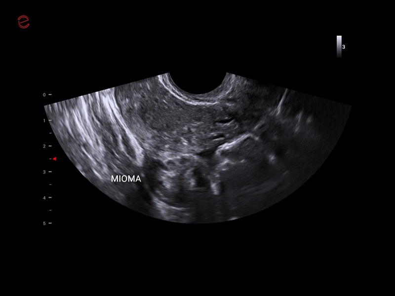
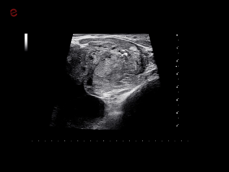
MyLab™X1 Go - WH Mioma
MyLab™X1 Go - WH Mioma
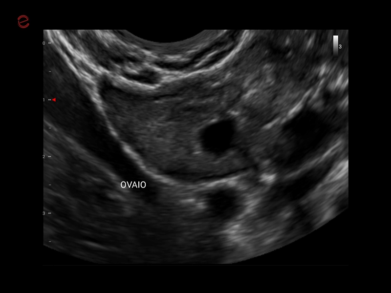
MyLab™X1 Go - WH Ovary
MyLab™X1 Go - WH Ovary
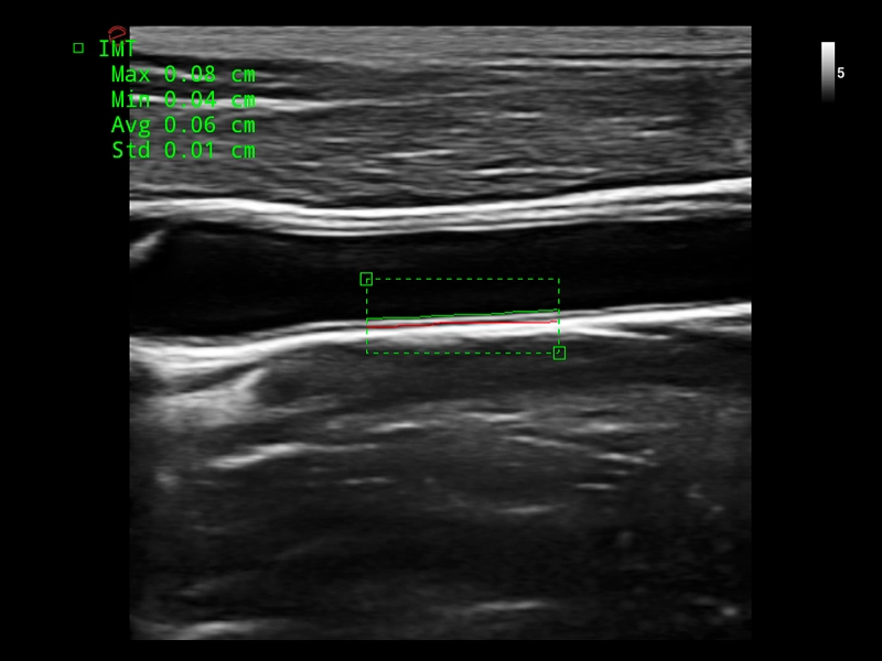
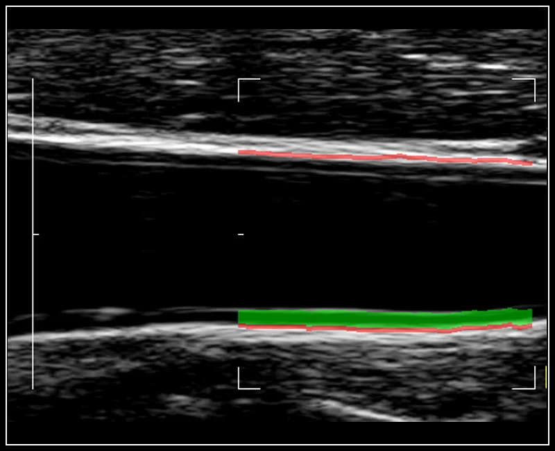
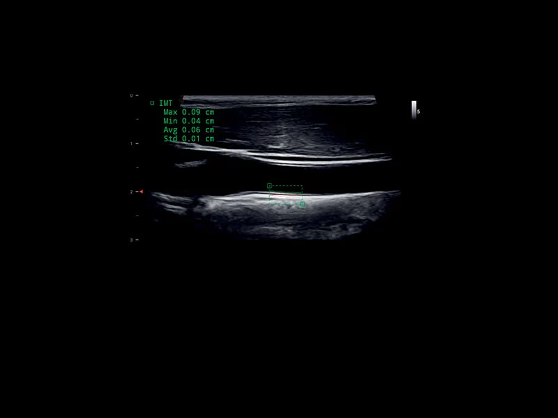
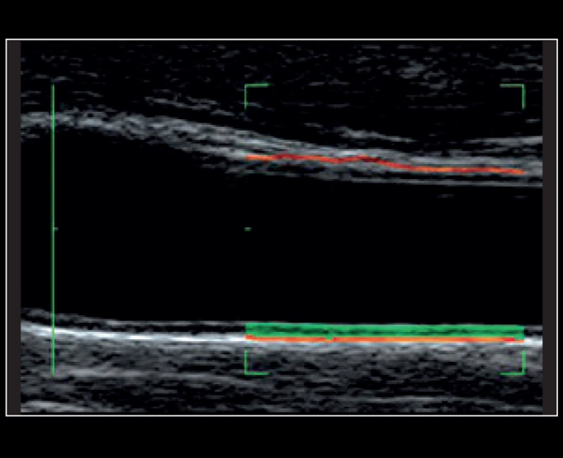
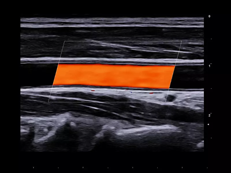
MyLab™X1 Go - Cardiovascular IMT
MyLab™X1 Go - Cardiovascular IMT
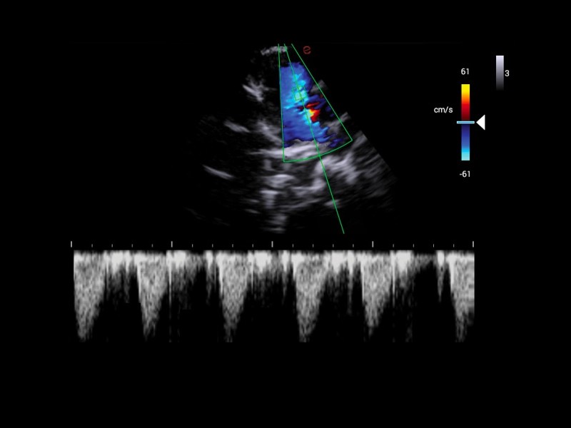
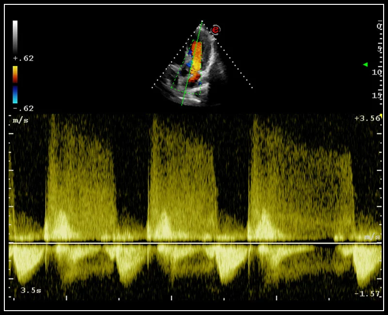
MyLab™X1 Go - Cardiovascular Doppler 02
MyLab™X1 Go - Cardiovascular Doppler 02
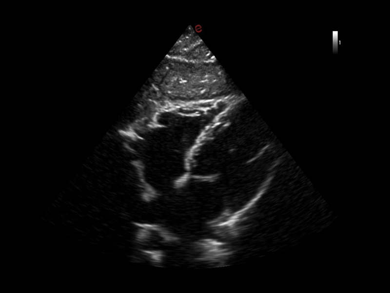
MyLab™X1 Go - Cardio fast
MyLab™X1 Go - Cardio fast
MyLab™X1 Go - Gyn
MyLab™X1 Go - Gyn
MyLab™X1 Go - Pelvic area
MyLab™X1 Go - Pelvic area
MyLab™X1 Go - Kidney
MyLab™X1 Go - Kidney
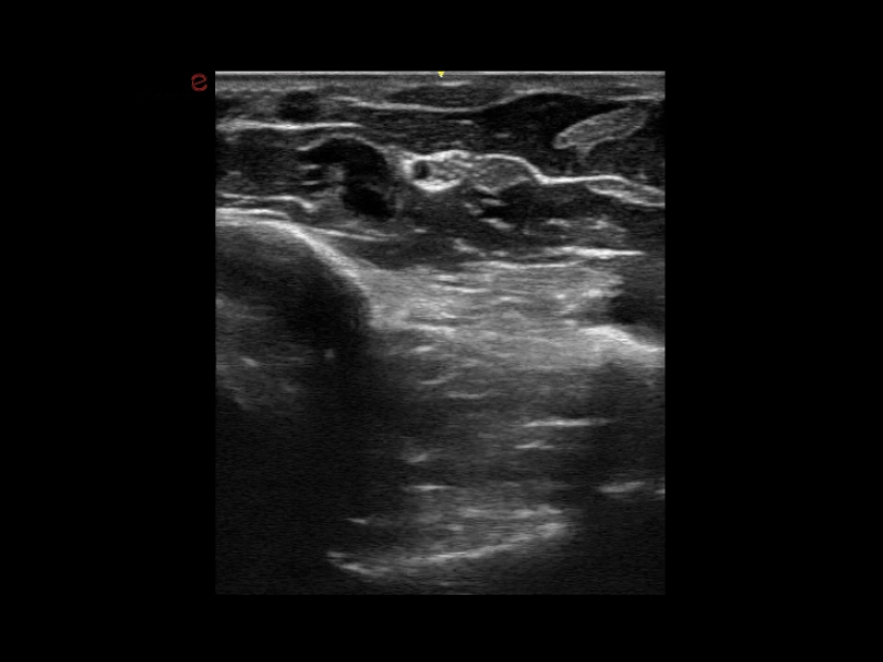
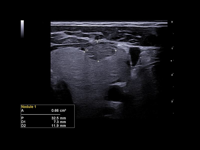
MyLab™X1 Go - Thyroid
MyLab™X1 Go - Thyroid
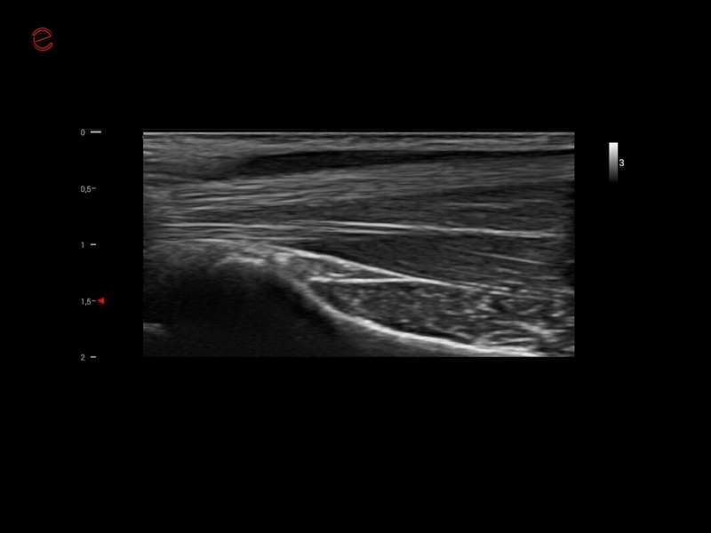
MyLab™X1 Go - Tendon
MyLab™X1 Go - Tendon
MyLab™X1 Go - Nerve
MyLab™X1 Go - Nerve
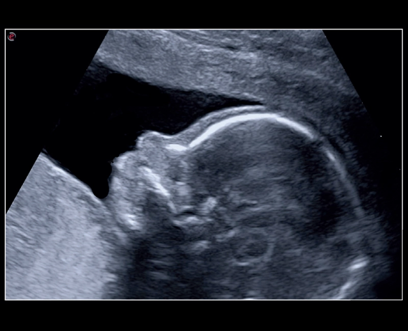
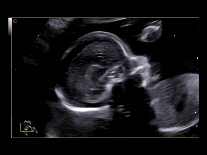
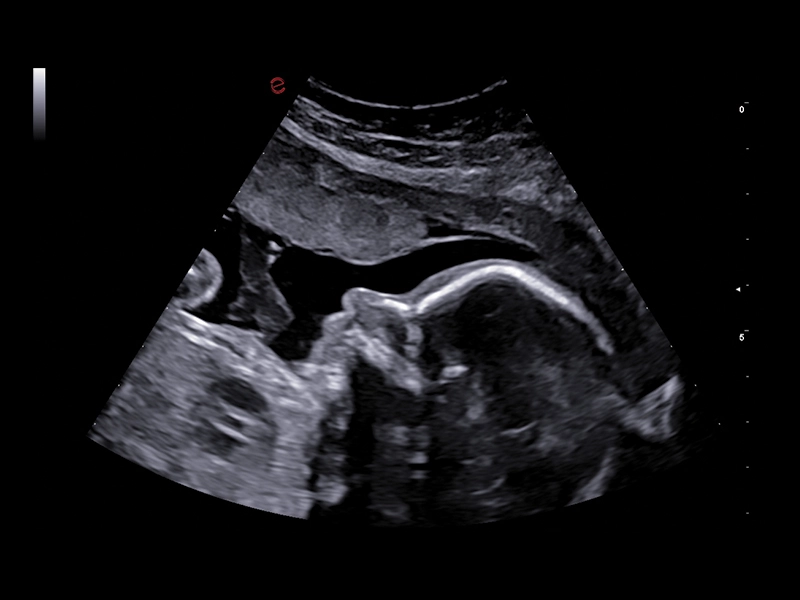
MyLab™E80 - B-Mode - Baby Profile
MyLab™E80 - B-Mode - Baby Profile
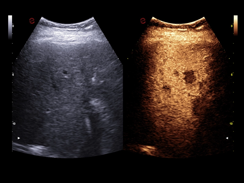
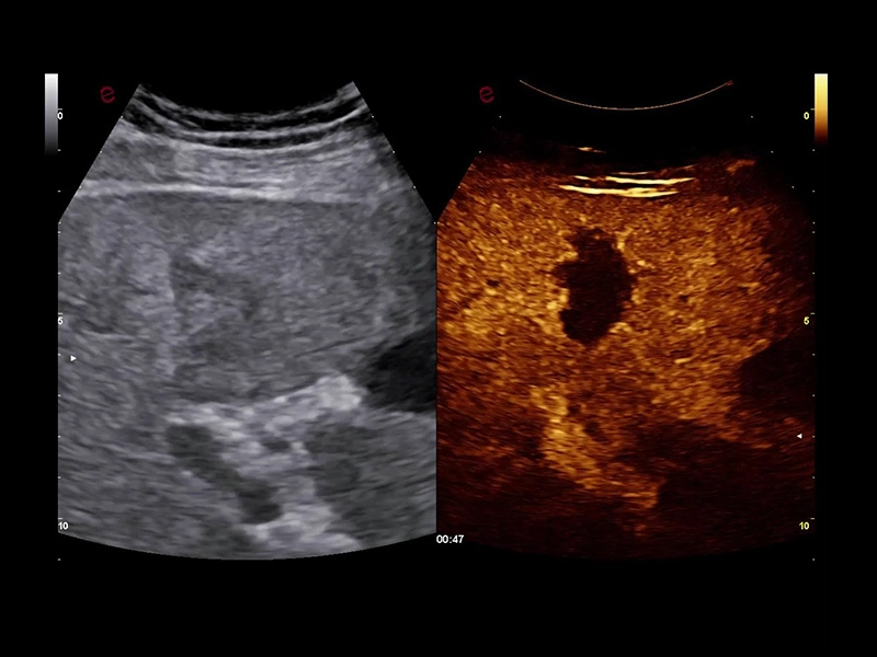
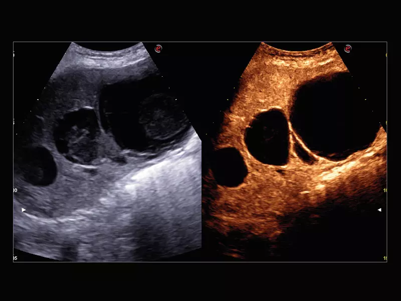
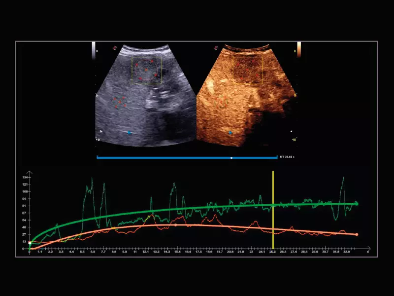
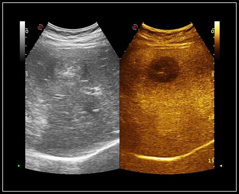
MyLab™E80 - CEUS - Liver
MyLab™E80 - CEUS - Liver
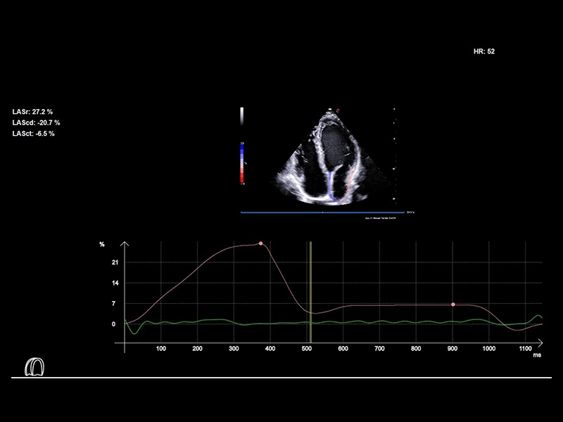
MyLab™E80 - XStrain LA
MyLab™E80 - XStrain LA
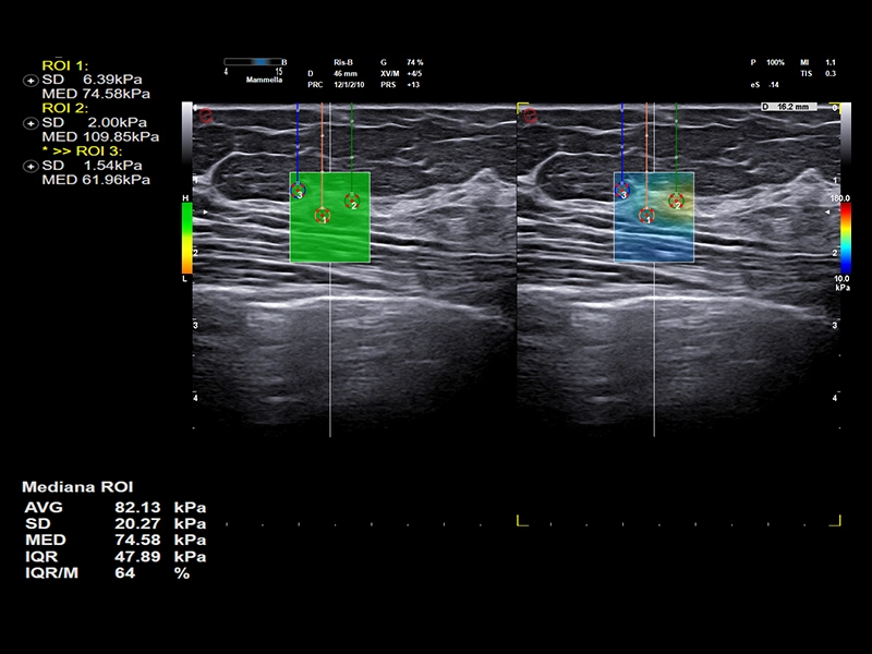
MyLab™E80 - QElaXto2D - Breast
MyLab™E80 - QElaXto2D - Breast
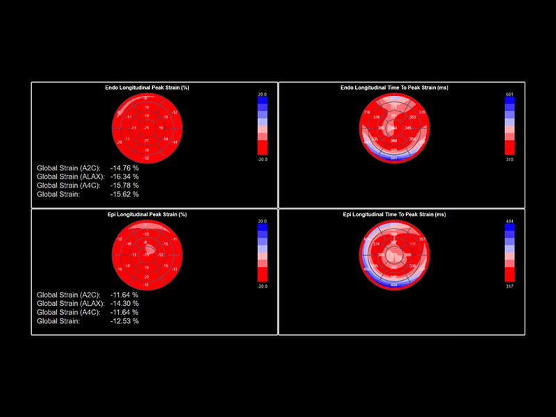
MyLab™E80 - XStrain LV - Bull's eye
MyLab™E80 - XStrain LV - Bull's eye
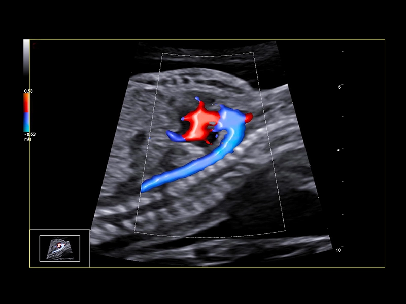
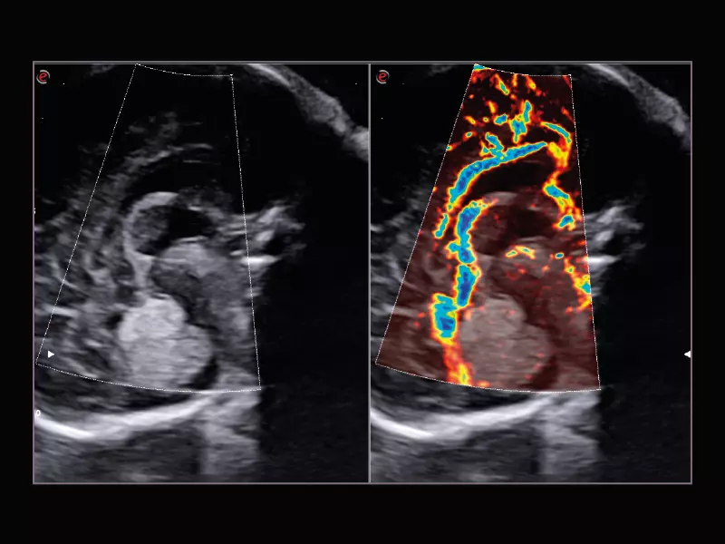
MyLab™E80 - BrightFlow - Aortic Arch
MyLab™E80 - BrightFlow - Aortic Arch
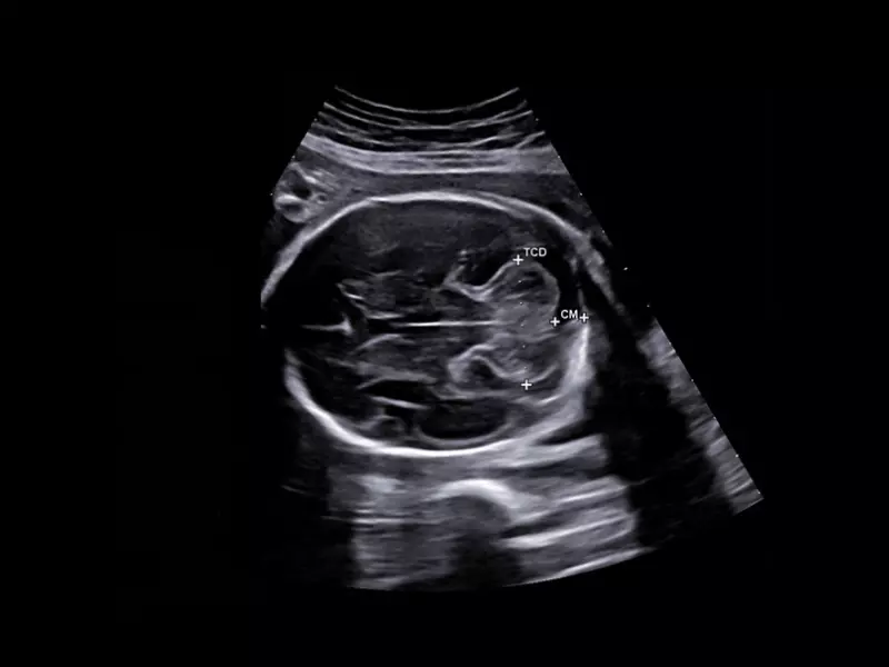
MyLab™E80 - AutoOB
MyLab™E80 - AutoOB
MyLab™E80 - VNav - Liver Fusion
MyLab™E80 - VNav - Liver Fusion
MyLab™E80 - QAI - Liver
MyLab™E80 - QAI - Liver
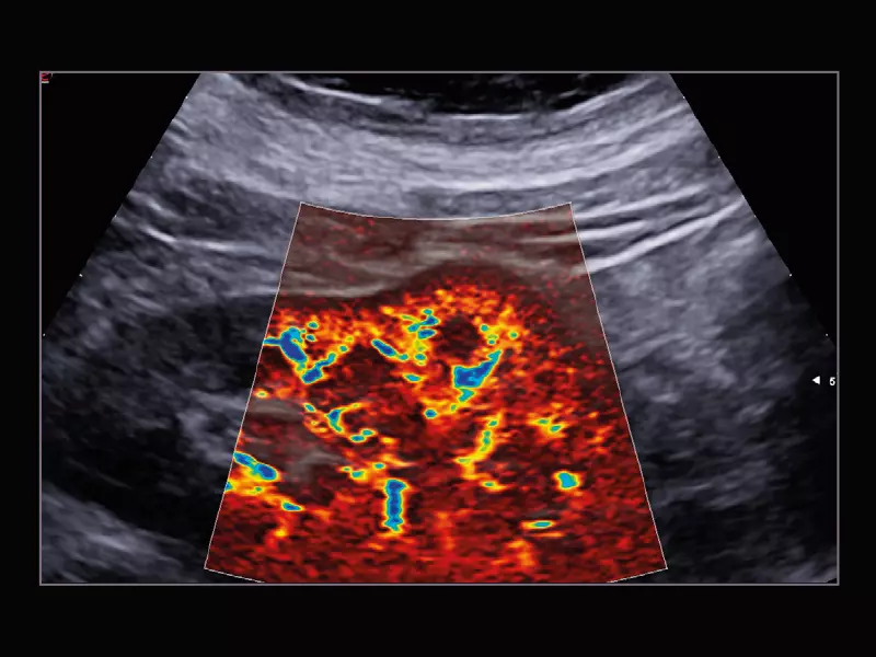
MyLab™E80 - microV - Kidney
MyLab™E80 - microV - Kidney
MyLab™E80 - LiverFusion
MyLab™E80 - LiverFusion
MyLab™E80 - B-Mode - Liver
MyLab™E80 - B-Mode - Liver
MyLab™E80 - Breast Interactive Workflow
MyLab™E80 - Breast Interactive Workflow
MyLab™E80 - Urofusion - TP Biopsy
MyLab™E80 - Urofusion - TP Biopsy
MyLab™E80 - Urofusion - Systematic Biopsy Guide
MyLab™E80 - Urofusion - Systematic Biopsy Guide
MyLab™E80 - 3D - Baby Face
MyLab™E80 - 3D - Baby Face
MyLab™E80 - microV - TCD
MyLab™E80 - microV - TCD
MyLab™E80 - HyperDoppler
MyLab™E80 - HyperDoppler

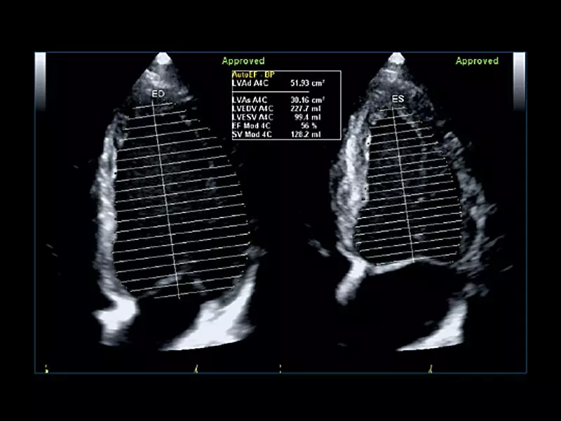
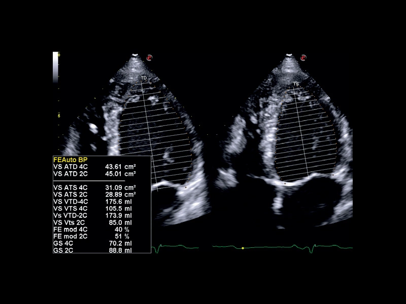
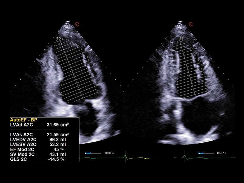
MyLab™E80 - AutoEF
MyLab™E80 - AutoEF
MyLab™E80 - AutoCM
MyLab™E80 - AutoCM
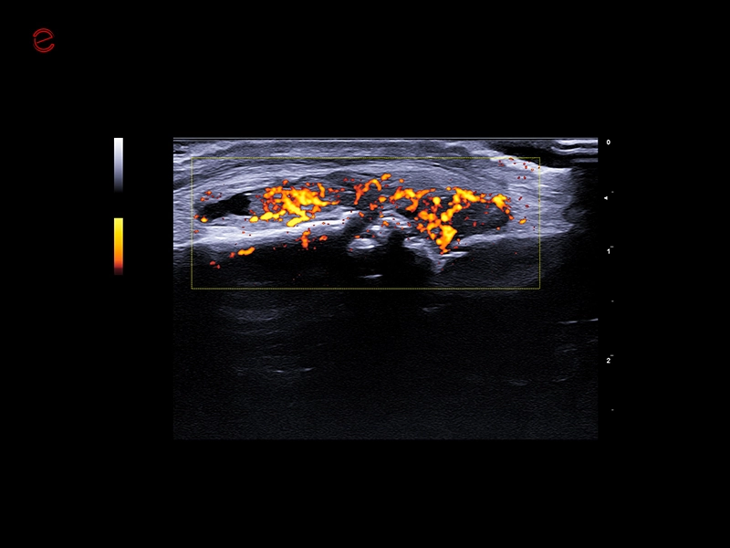
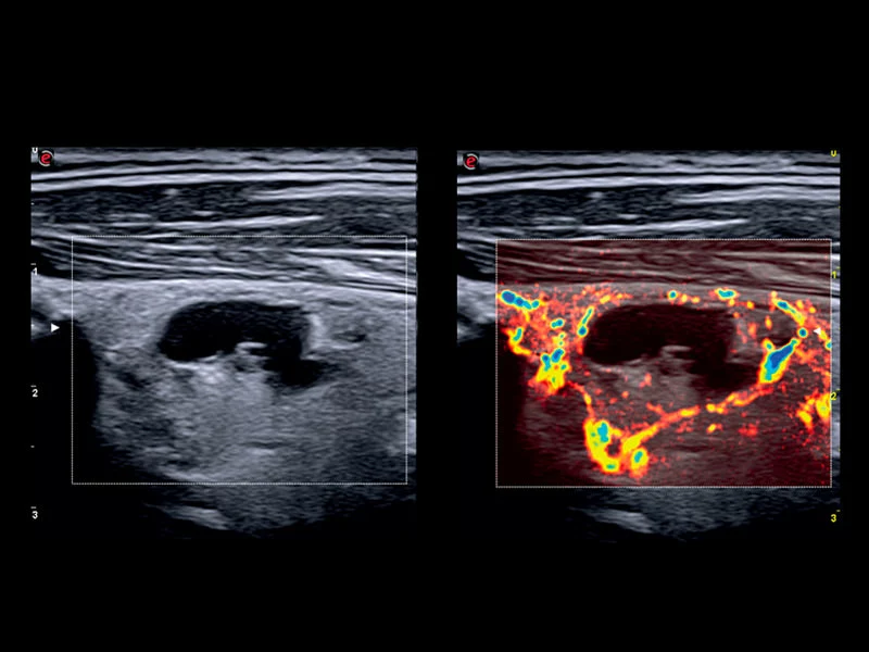
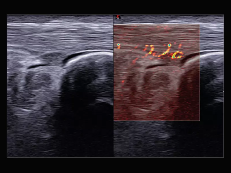
MyLab™E80 - microV - Thyroid
MyLab™E80 - microV - Thyroid
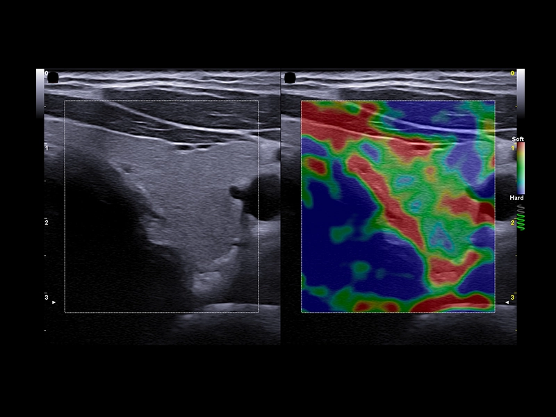
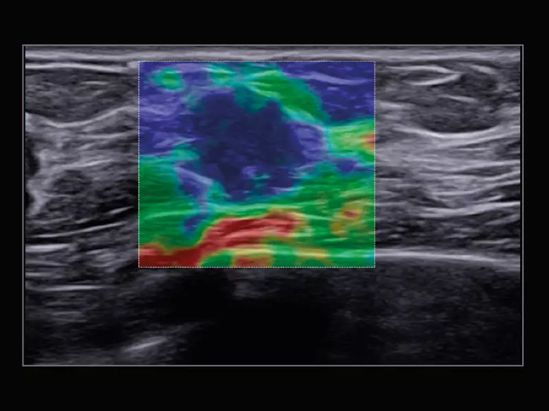
MyLab™E80 - ElaXto - Thyroid
MyLab™E80 - ElaXto - Thyroid
MyLab™E80 - eDetect - Thyroid
MyLab™E80 - eDetect - Thyroid
MyLab™E80 - eDetect - Breast
MyLab™E80 - eDetect - Breast
MyLab™E80 - LX 3-15 - B-Mode - MSK
MyLab™E80 - LX 3-15 - B-Mode - MSK
MyLab™E80 - microV - MSK
MyLab™E80 - microV - MSK
MyLab™E80 - B-Mode - Nerve
MyLab™E80 - B-Mode - Nerve
MyLab™E80 - B-Mode - MSK
MyLab™E80 - B-Mode - MSK
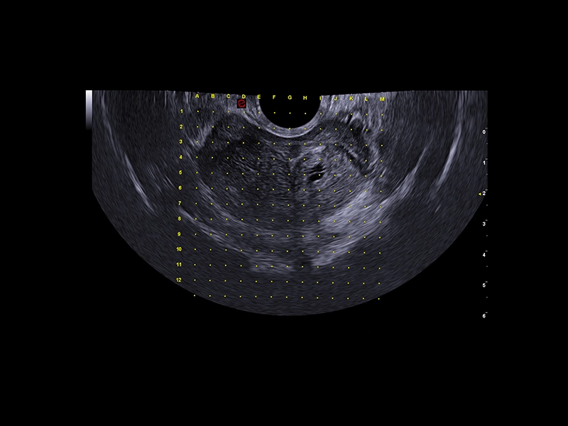
MyLab™E80 - Urofusion - Sample Mapping
MyLab™E80 - Urofusion - Sample Mapping
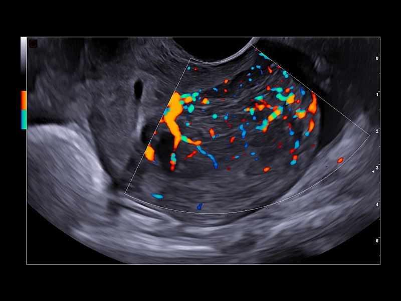
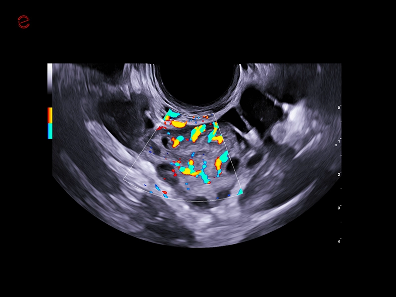
MyLab™E80 - Power Doppler - Gynaecology
MyLab™E80 - Power Doppler - Gynaecology
MyLab™E80 - Classic CIVCO Stepper
MyLab™E80 - Classic CIVCO Stepper
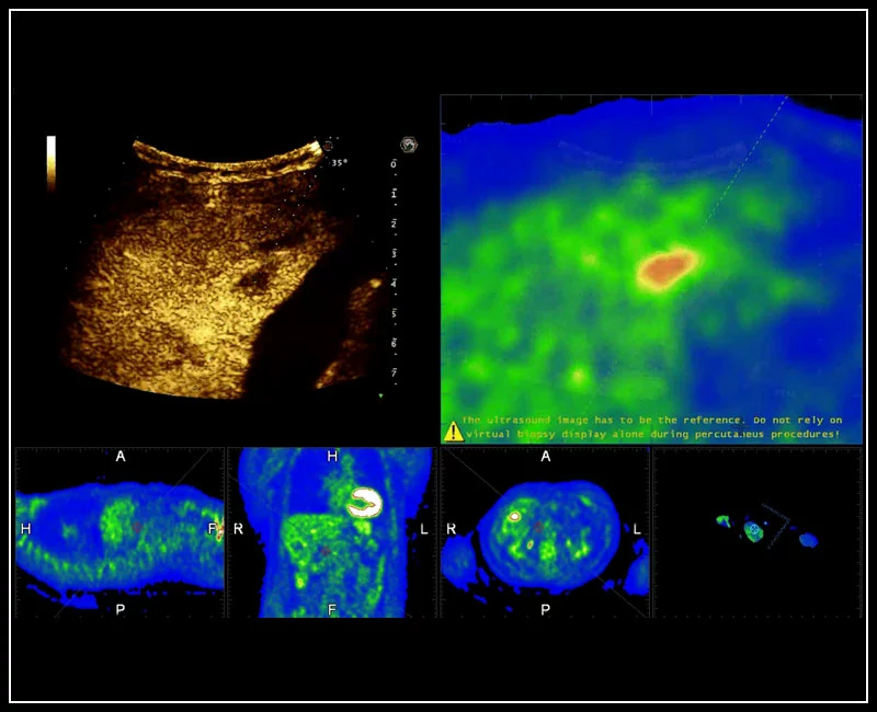
MyLab™9 Platform - Real-time CEUS and PET fusion for lesion detection
MyLab™9 Platform - Real-time CEUS and PET fusion for lesion detection
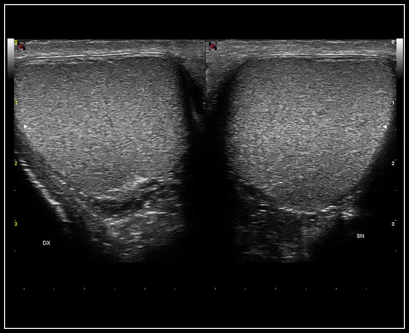
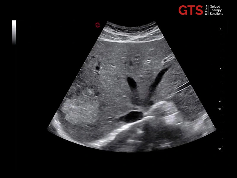
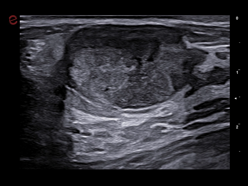
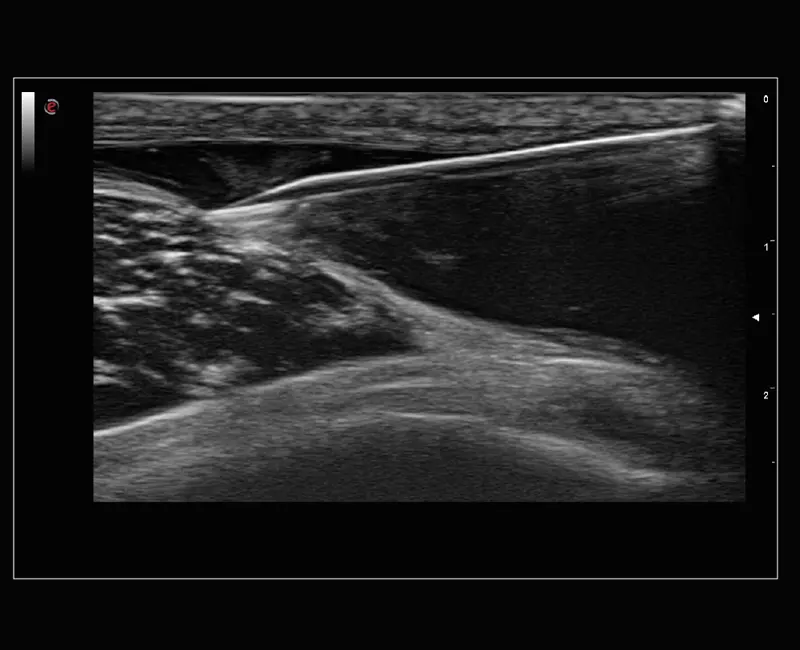
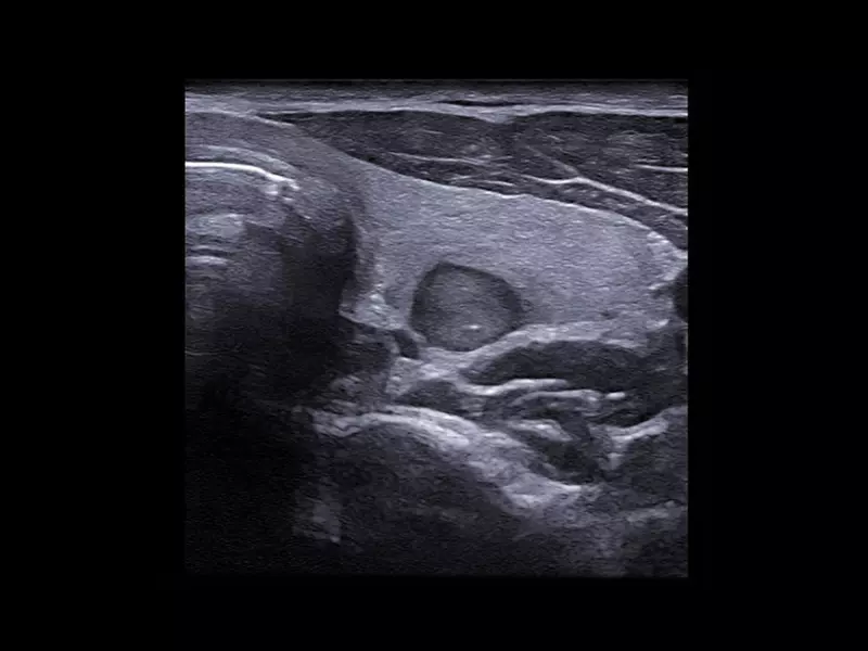
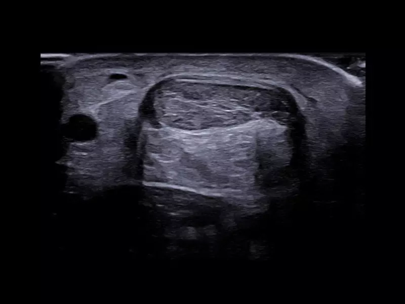
MyLab™9 Platform - High resolution imaging in testis
MyLab™9 Platform - High resolution imaging in testis
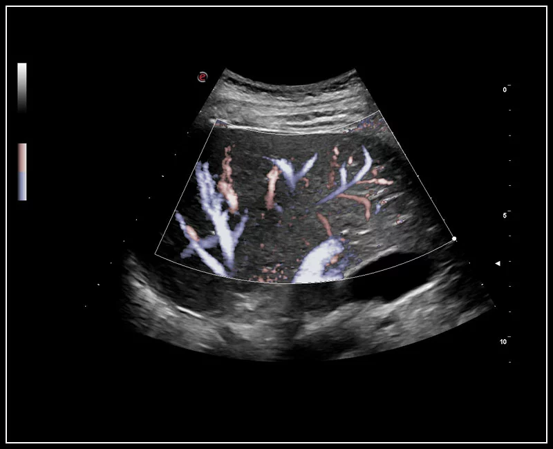
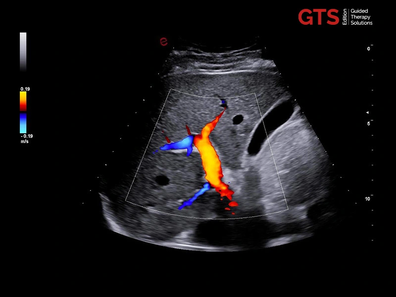
MyLab™9 Platform - XFlow Doppler enhancement in liver vascularization
MyLab™9 Platform - XFlow Doppler enhancement in liver vascularization
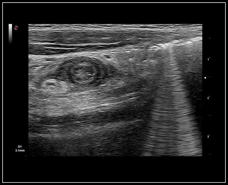
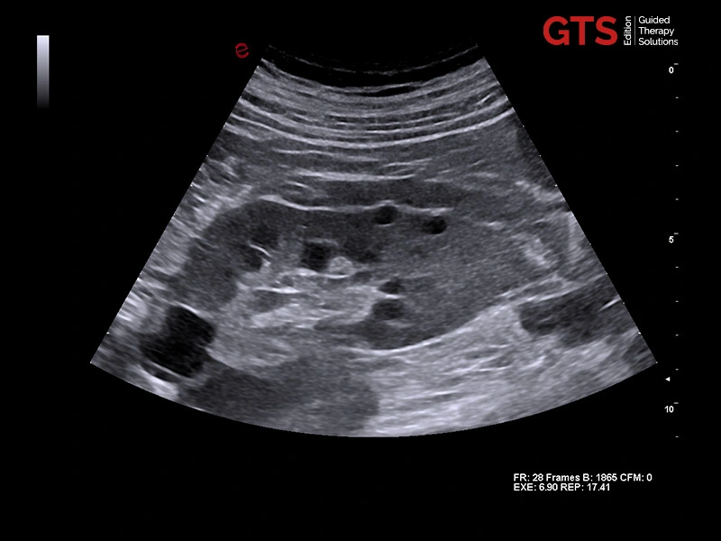
MyLab™9 Platform - Axial view of appendicitis
MyLab™9 Platform - Axial view of appendicitis
MyLab™9 Platform - Dual ElaXto characterization on breast lesion
MyLab™9 Platform - Dual ElaXto characterization on breast lesion
MyLab™9 Platform - QPack quantification capabilities on-board even with CFM
MyLab™9 Platform - QPack quantification capabilities on-board even with CFM
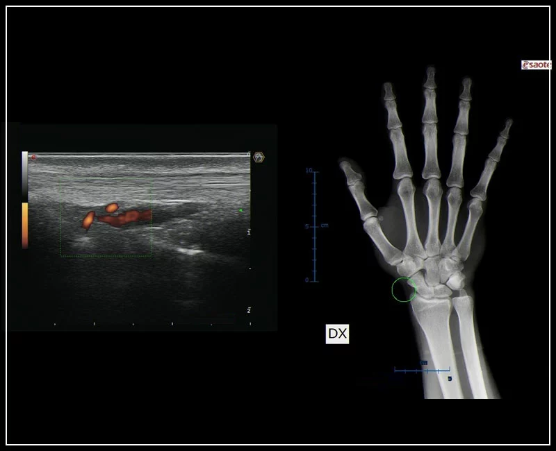
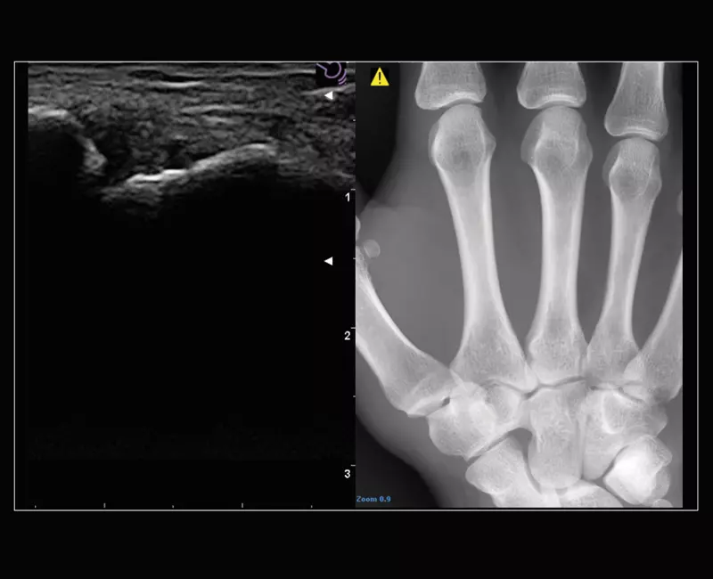
MyLab™9 Platform - MSK BodyMap and real-time XFlow on X-Ray extremities
MyLab™9 Platform - MSK BodyMap and real-time XFlow on X-Ray extremities
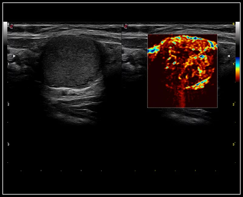
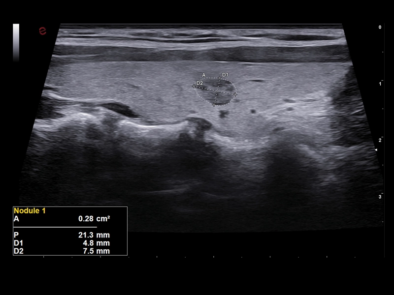
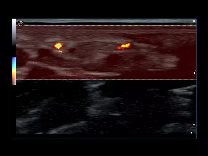
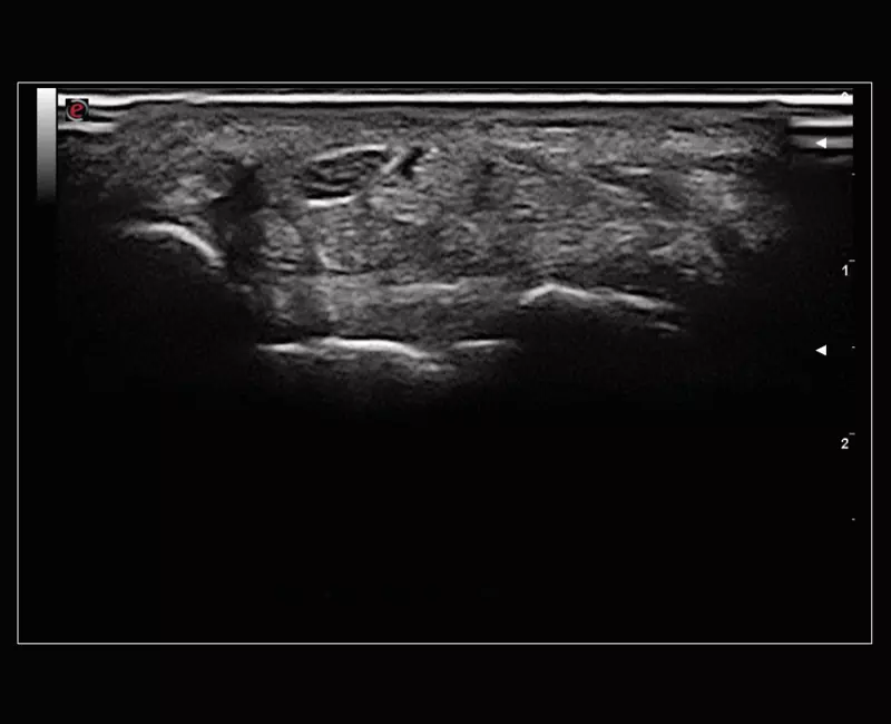
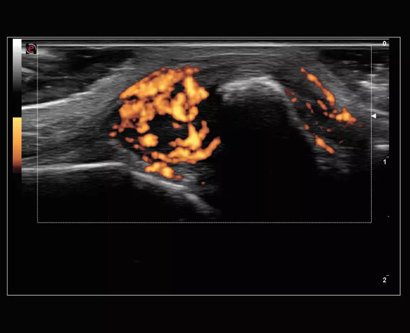
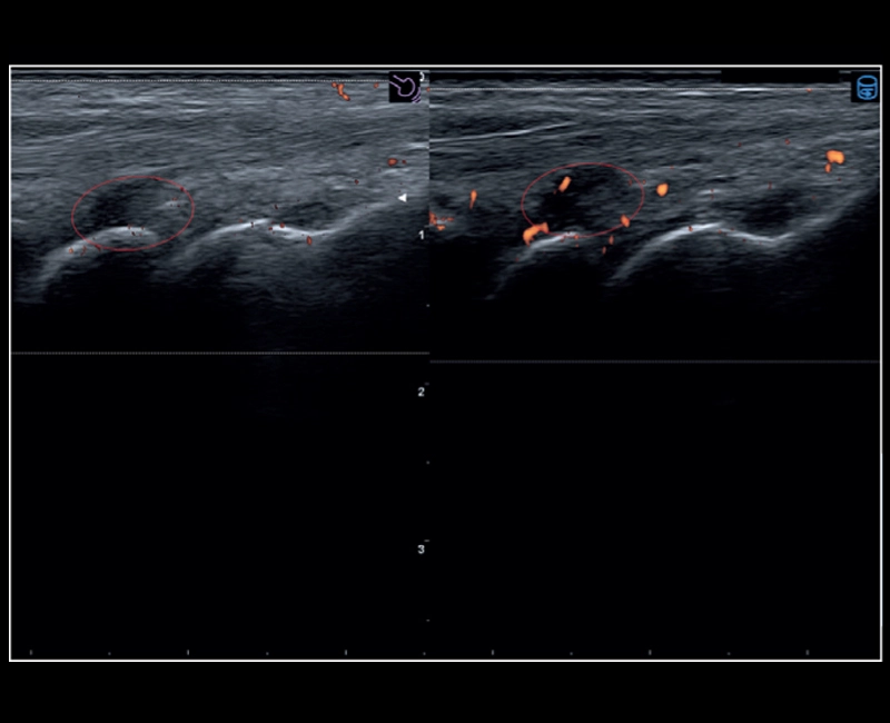
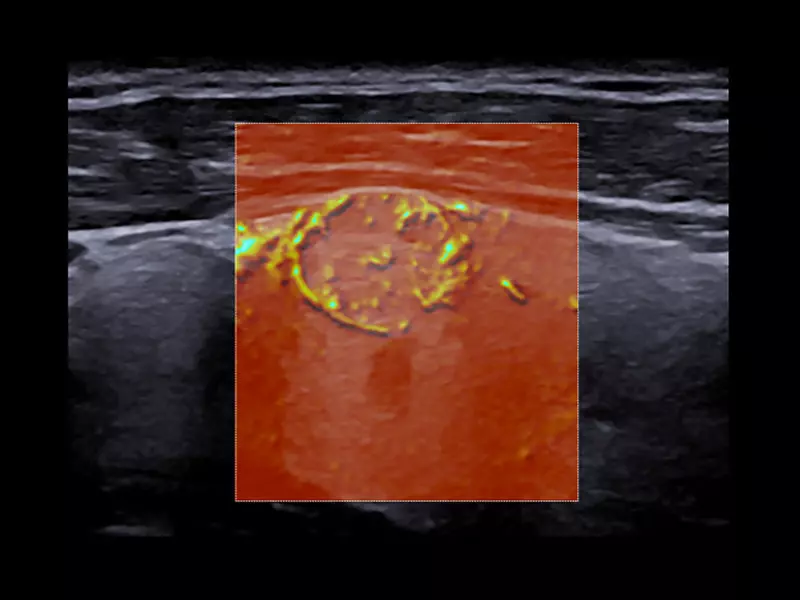
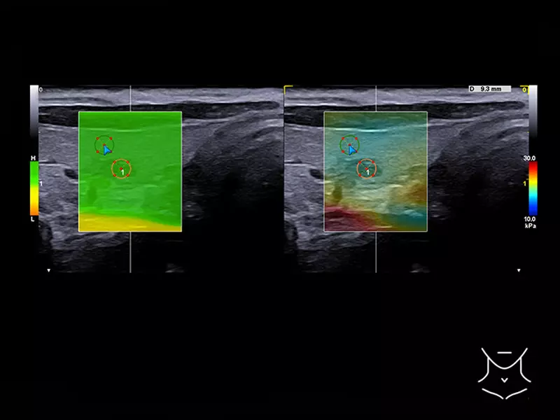
MyLab™9 Platform - Advanced hemodynamic analysis in thyroid nodule with microV
MyLab™9 Platform - Advanced hemodynamic analysis in thyroid nodule with microV
MyLab™9 Platform - QElaXto point Shearwave Elastography in liver
MyLab™9 Platform - QElaXto point Shearwave Elastography in liver
MyLab™9 Platform - Pre-Post volumetric CEUS-multidataset comparison
MyLab™9 Platform - Pre-Post volumetric CEUS-multidataset comparison
MyLab™9 Platform - Breast BodyMap and real-time ElaXto in Mammo
MyLab™9 Platform - Breast BodyMap and real-time ElaXto in Mammo
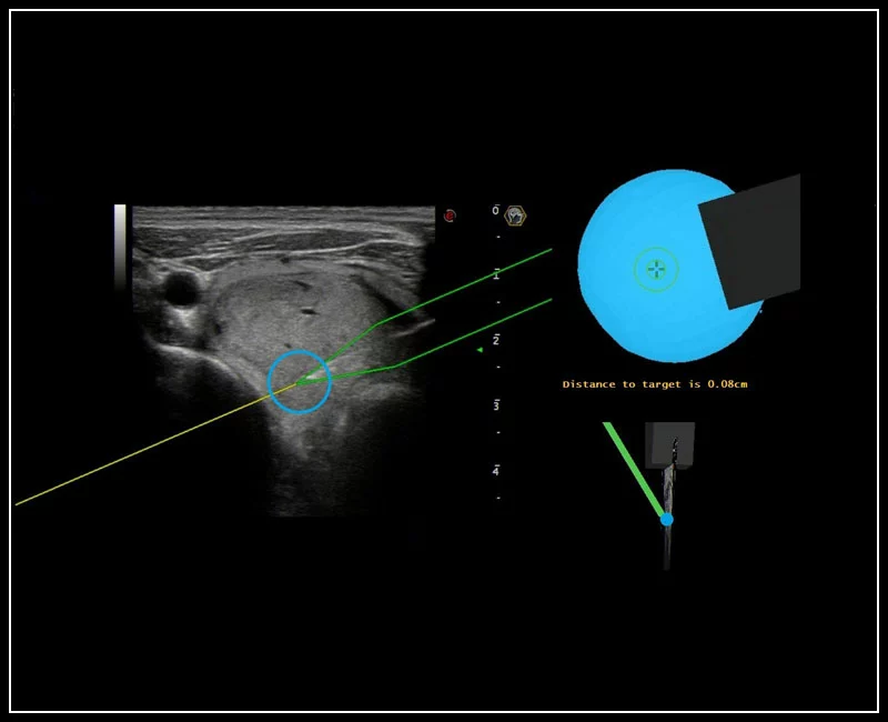
MyLab™9 Platform - Precise lesion detection and guidance with Virtual Biopsy
MyLab™9 Platform - Precise lesion detection and guidance with Virtual Biopsy
MyLab™9 Platform - Virtual Navigator automatic vascular detection and segmentation with Auto-Fusion
MyLab™9 Platform - Virtual Navigator automatic vascular detection and segmentation with Auto-Fusion
MyLab™9 Platform - Gynecology fusion imaging with PET for best lesion location
MyLab™9 Platform - Gynecology fusion imaging with PET for best lesion location
MyLab™9 Platform - easyTrace to maximize Doppler performance
MyLab™9 Platform - easyTrace to maximize Doppler performance
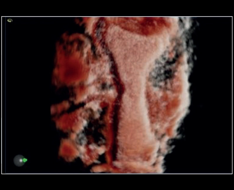
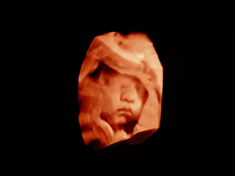
MyLab™9 Platform - Real-time baby face with 4D imaging
MyLab™9 Platform - Real-time baby face with 4D imaging
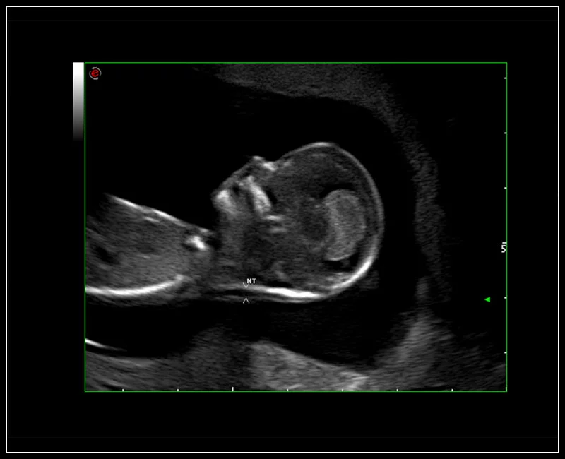
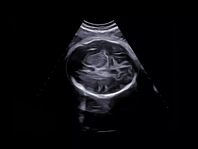
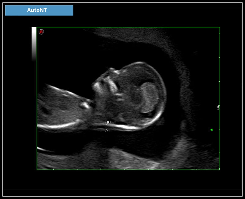
MyLab™9 Platform - HD Zoom on fetus profile with AutoNT measurement
MyLab™9 Platform - HD Zoom on fetus profile with AutoNT measurement
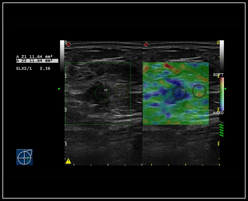
MyLab™9 Platform - Elastography advanced measurement package on breast lesion
MyLab™9 Platform - Elastography advanced measurement package on breast lesion
MyLab™9 Platform - 24MHz Imaging even on fingerprint with CFM
MyLab™9 Platform - 24MHz Imaging even on fingerprint with CFM
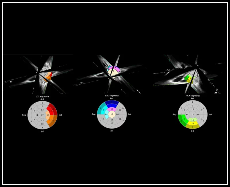
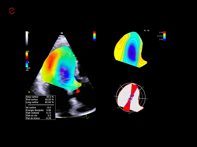
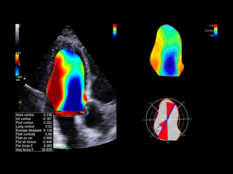
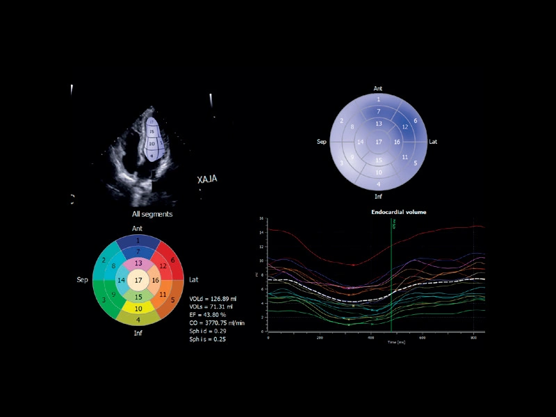
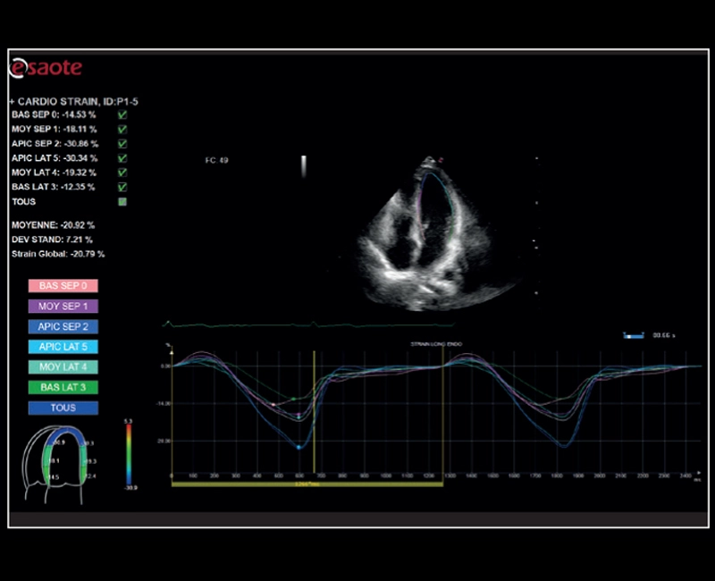
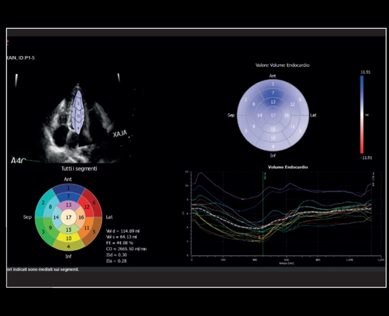
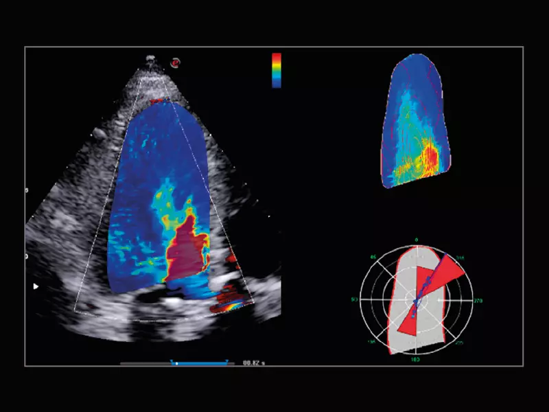
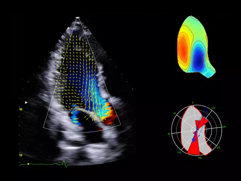
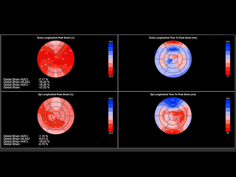
MyLab™9 Platform - XStrain4D technology for volumetric heart assessment by coronary territories
MyLab™9 Platform - XStrain4D technology for volumetric heart assessment by coronary territories
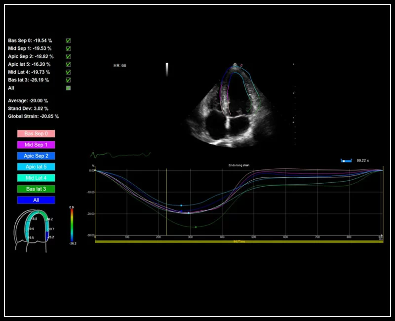
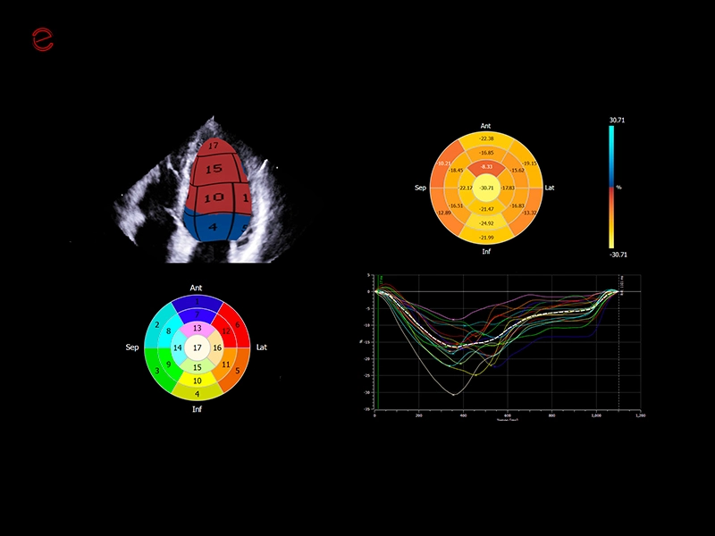
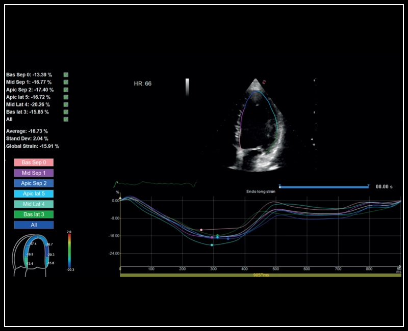
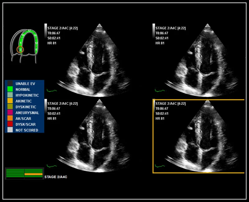
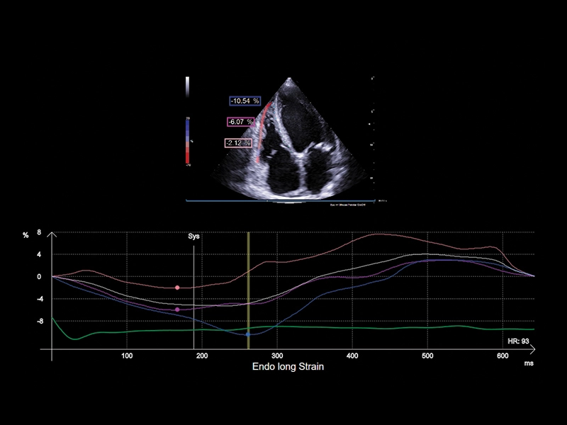
MyLab™9 Platform - XStrain™ 2D speckle tracking technologies for global and regional function
MyLab™9 Platform - XStrain™ 2D speckle tracking technologies for global and regional function
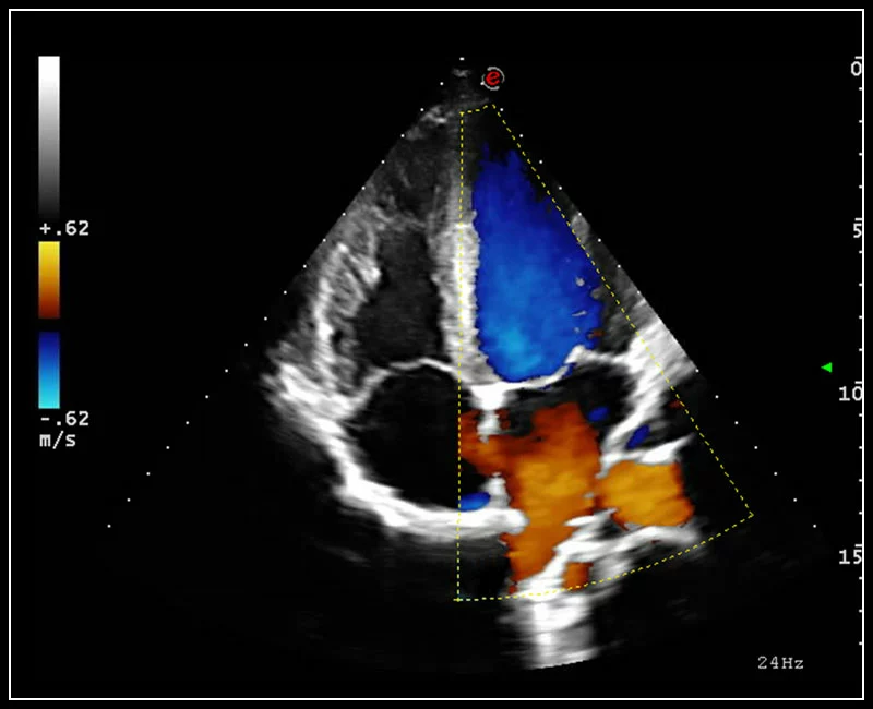
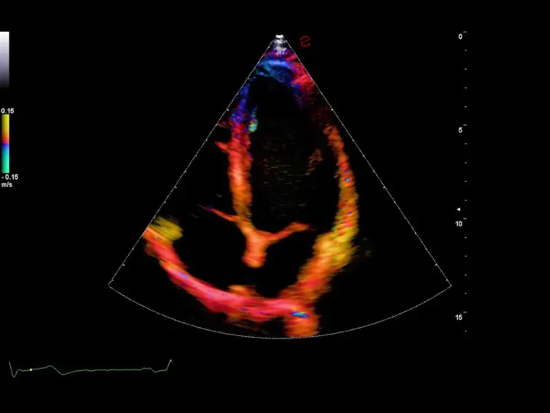
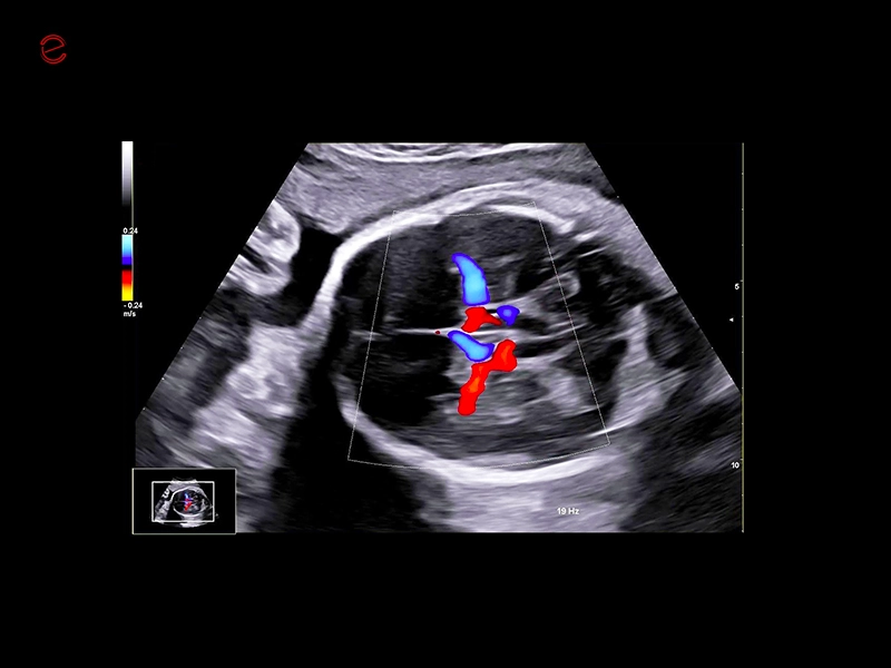
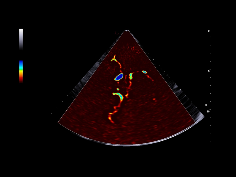
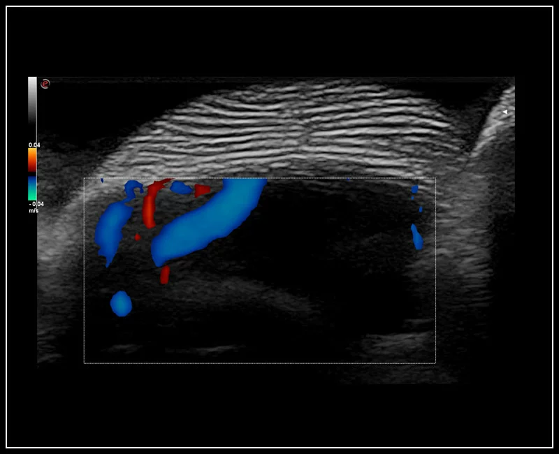
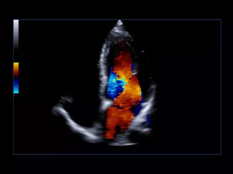
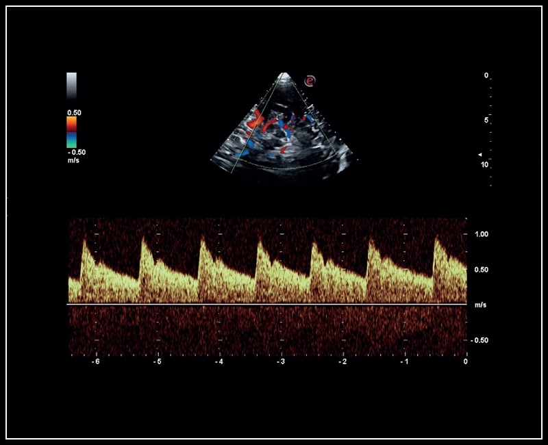
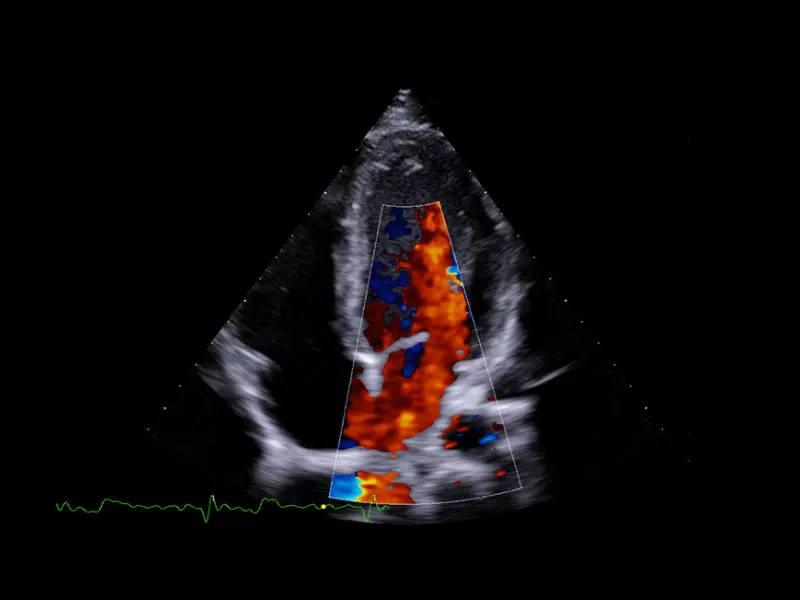
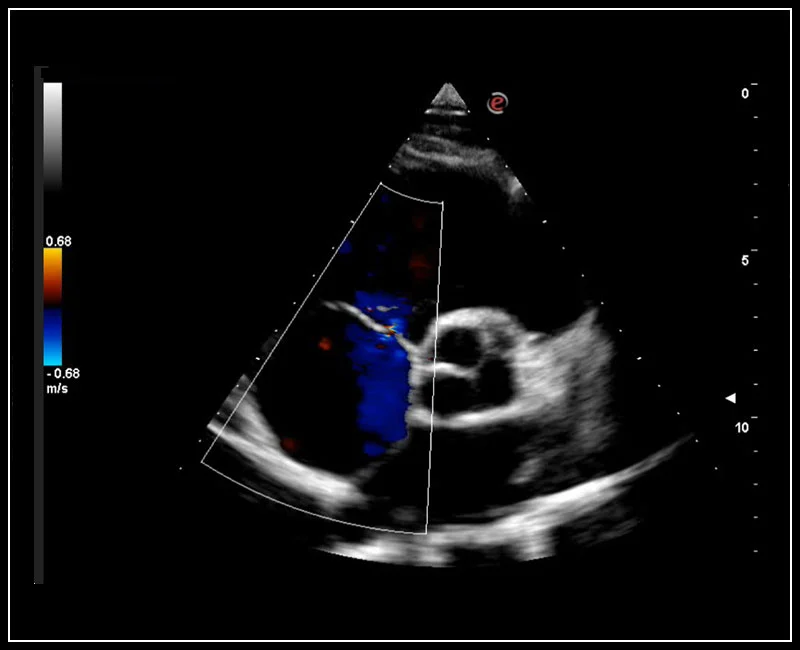
MyLab™9 Platform - Ultra-sensitivity Colour Doppler for precise visualization pulmonary veins
MyLab™9 Platform - Ultra-sensitivity Colour Doppler for precise visualization pulmonary veins
MyLab™9 Platform - XView real-time algorithm reducing speckle noise artefact in pathologies analysis quantification
MyLab™9 Platform - XView real-time algorithm reducing speckle noise artefact in pathologies analysis quantification
MyLab™9 Platform - High Frequency MSK Imaging with HD Zoom
MyLab™9 Platform - High Frequency MSK Imaging with HD Zoom
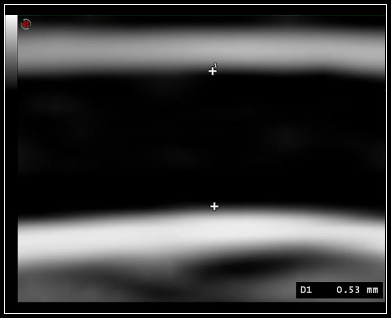
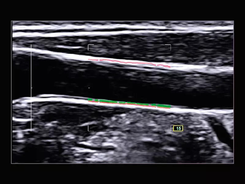
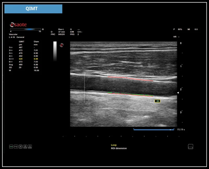
MyLab™9 Platform - QIMT Intima-media thickness quantification based on radio frequencies in real-time studies
MyLab™9 Platform - QIMT Intima-media thickness quantification based on radio frequencies in real-time studies
MyLab™9 Platform - Advanced CW Doppler processing chain for aortic stenosis quantification
MyLab™9 Platform - Advanced CW Doppler processing chain for aortic stenosis quantification
MyLab™9 Platform - Tissue Velocity imaging to quantify septal velocity and dyssynchrony
MyLab™9 Platform - Tissue Velocity imaging to quantify septal velocity and dyssynchrony
MyLab™9 Platform - Very-superficial linear imaging with Power Doppler algorithm
MyLab™9 Platform - Very-superficial linear imaging with Power Doppler algorithm
MyLab™9 Platform - PW Doppler with easyTrace optimization
MyLab™9 Platform - PW Doppler with easyTrace optimization
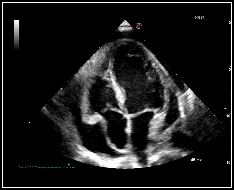
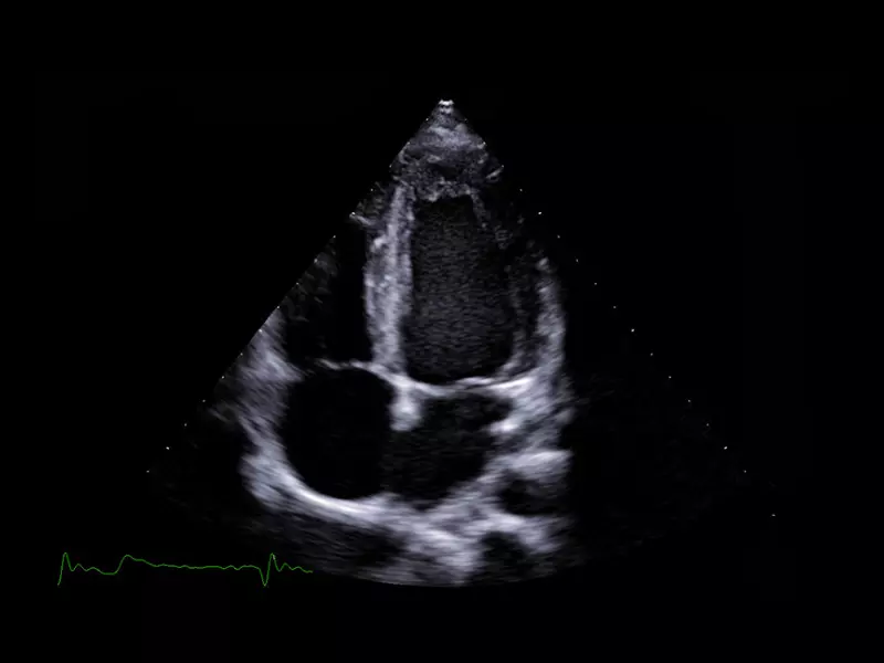
MyLab™9 Platform - TEI™ harmonic imaging for clear visualization of perimembranous ventricular septal
MyLab™9 Platform - TEI™ harmonic imaging for clear visualization of perimembranous ventricular septal
MyLab™9 Platform - Ultra-sensitivity Colour Doppler in detection vertebral artery
MyLab™9 Platform - Ultra-sensitivity Colour Doppler in detection vertebral artery
MyLab™9 Platform - HD zoom transcranial vascularization analysis with microV
MyLab™9 Platform - HD zoom transcranial vascularization analysis with microV
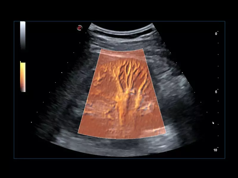
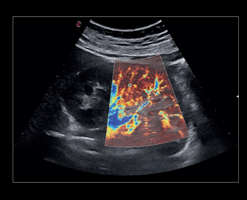
MyLab™9 Platform - Hemodynamics analysis using microV on kidney
MyLab™9 Platform - Hemodynamics analysis using microV on kidney
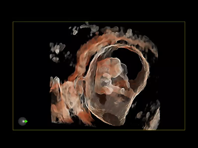
MyLab™9 Platform - Baby Face rendering in real-time with XLight
MyLab™9 Platform - Baby Face rendering in real-time with XLight
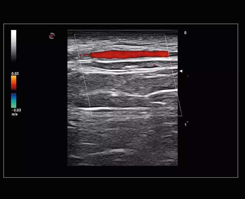
MyLab™9 Platform - XFlow in testis vascularization
MyLab™9 Platform - XFlow in testis vascularization
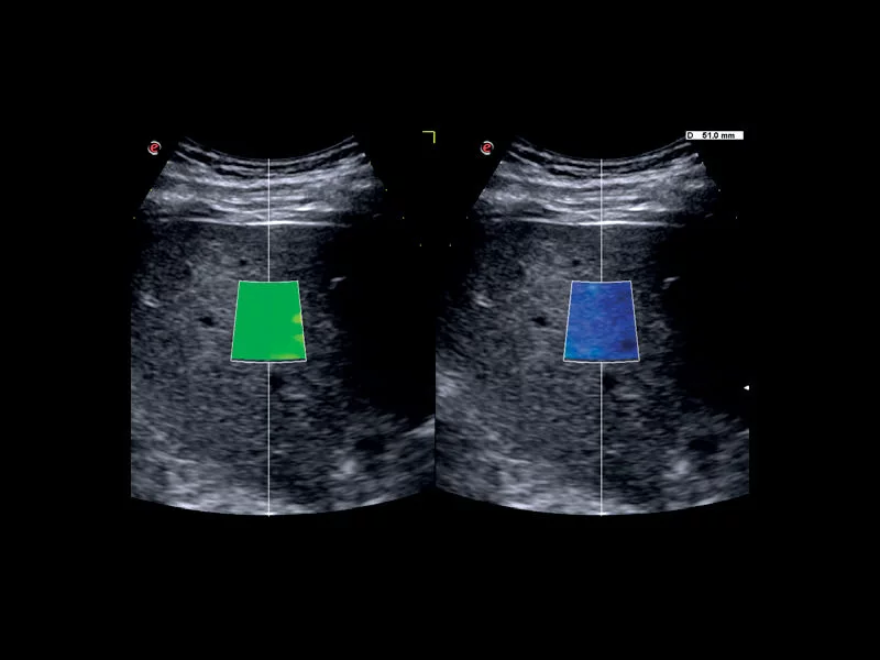
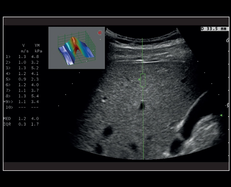
MyLab™9 Platform - QElaXto 2D shearwave elastography in liver
MyLab™9 Platform - QElaXto 2D shearwave elastography in liver
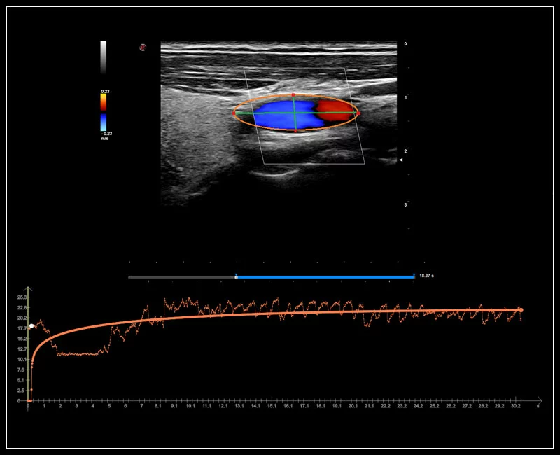
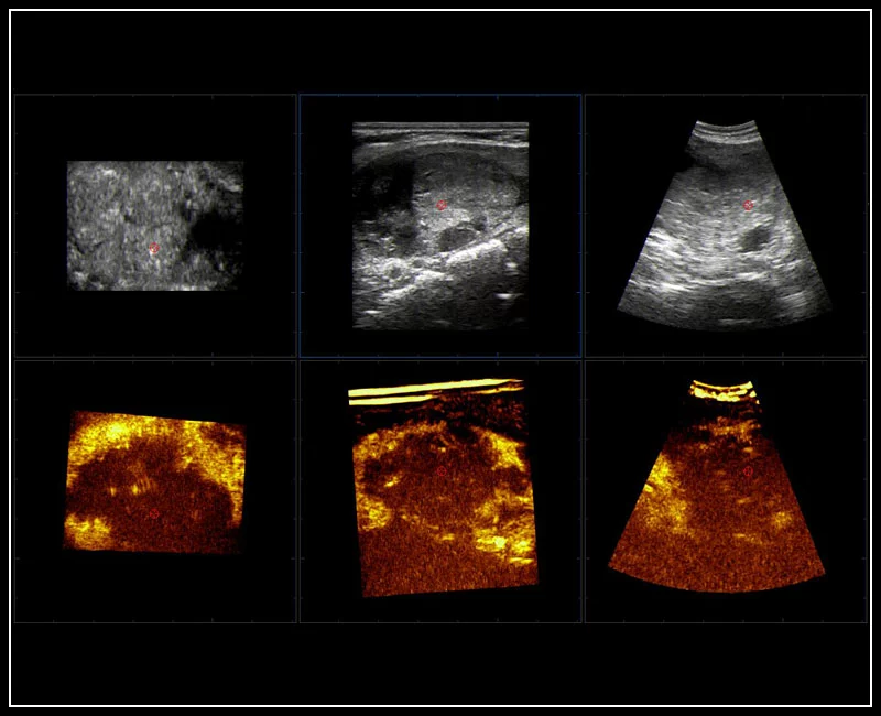
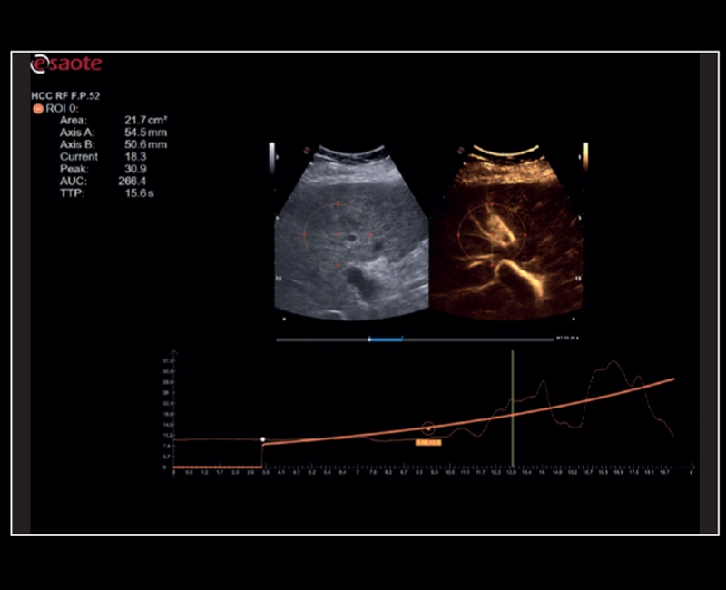
MyLab™9 Platform - QPack CnTI™ perfusion analysis post RF ablation
MyLab™9 Platform - QPack CnTI™ perfusion analysis post RF ablation
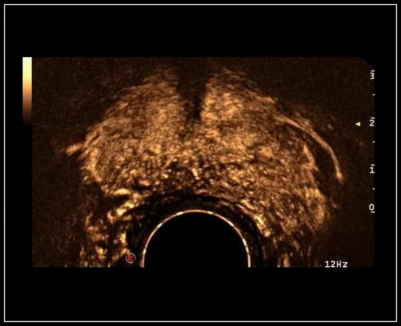
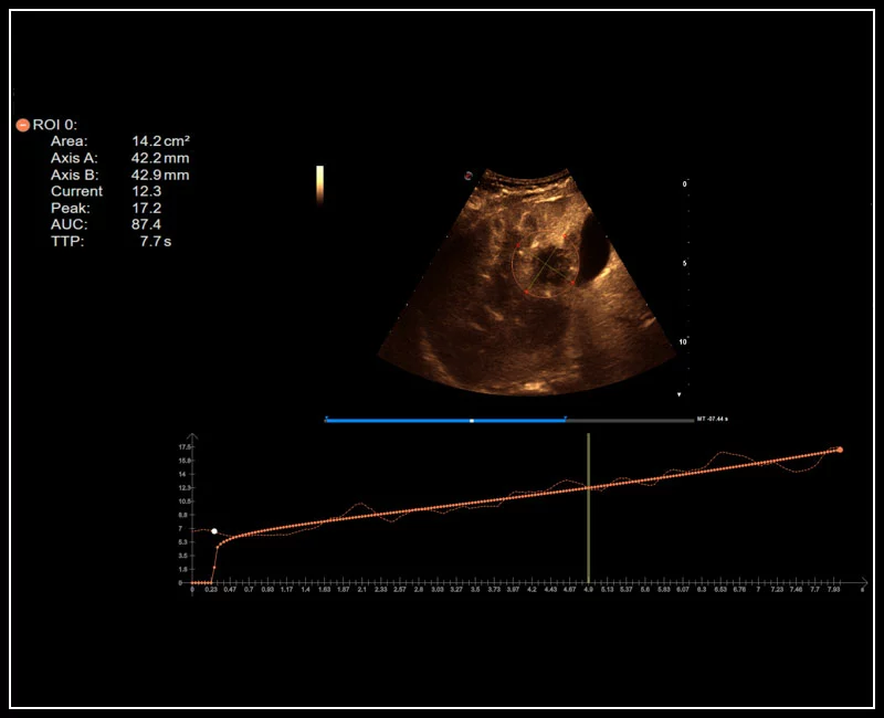
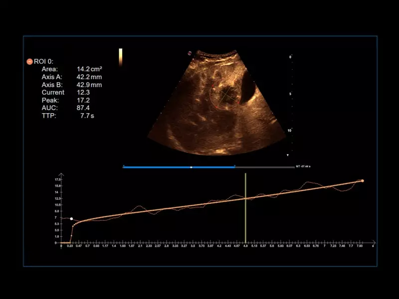
MyLab™9 Platform - Prostate contrast enhanced imaging (CnTI™)
MyLab™9 Platform - Prostate contrast enhanced imaging (CnTI™)
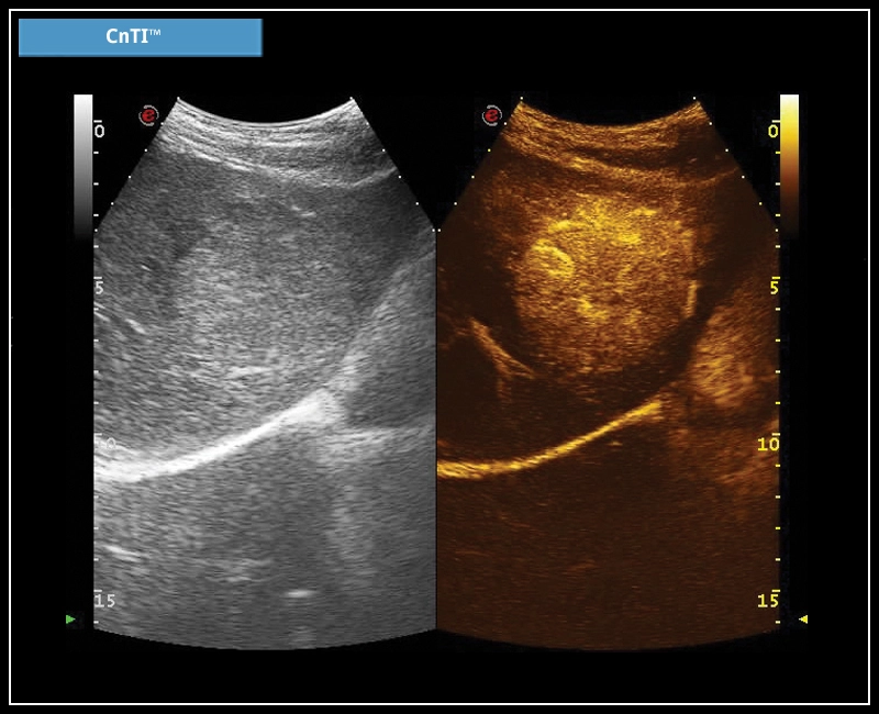
MyLab™Omega eXP - CnTI™
MyLab™Omega eXP - CnTI™
MyLab™Omega eXP - Cardiovascular 01
MyLab™Omega eXP - Cardiovascular 01
MyLab™Omega eXP - VPan
MyLab™Omega eXP - VPan
MyLab™Omega eXP - General Imaging 01
MyLab™Omega eXP - General Imaging 01
MyLab™Omega eXP - eDetect
MyLab™Omega eXP - eDetect
MyLab™Omega eXP - Women's Health 01
MyLab™Omega eXP - Women's Health 01
MyLab™Omega eXP - AutoOB
MyLab™Omega eXP - AutoOB
MyLab™Omega eXP - General Imaging 02
MyLab™Omega eXP - General Imaging 02
MyLab™Omega eXP - BrightFlow
MyLab™Omega eXP - BrightFlow
MyLab™Omega eXP - Cardiovascular 02
MyLab™Omega eXP - Cardiovascular 02
MyLab™Omega eXP -microV
MyLab™Omega eXP -microV
MyLab™Omega eXP - General Imaging 03
MyLab™Omega eXP - General Imaging 03
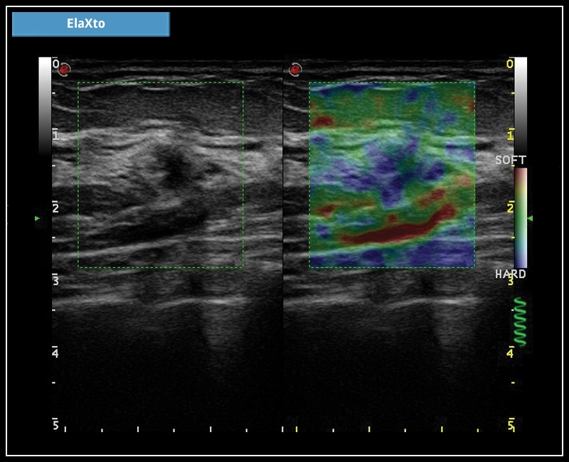
MyLab™Omega eXP - ElaXto
MyLab™Omega eXP - ElaXto
MyLab™Omega eXP - Women's Health 02
MyLab™Omega eXP - Women's Health 02
MyLab™Omega eXP - XGlass
MyLab™Omega eXP - XGlass
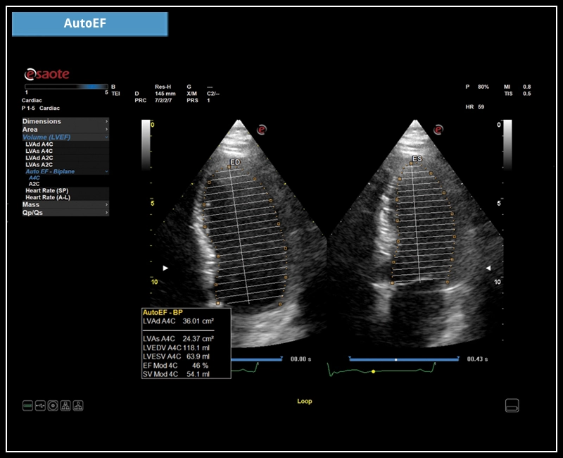
MyLab™Omega eXP - AutoEF
MyLab™Omega eXP - AutoEF
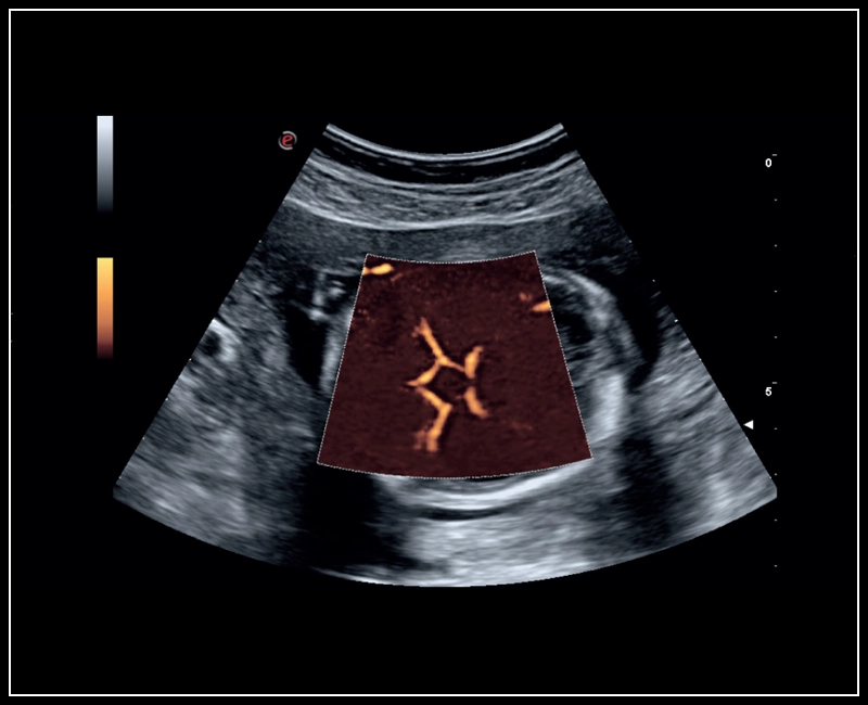
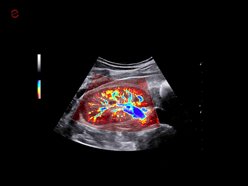
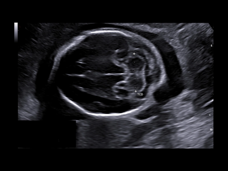
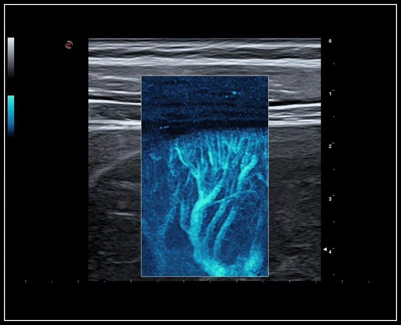
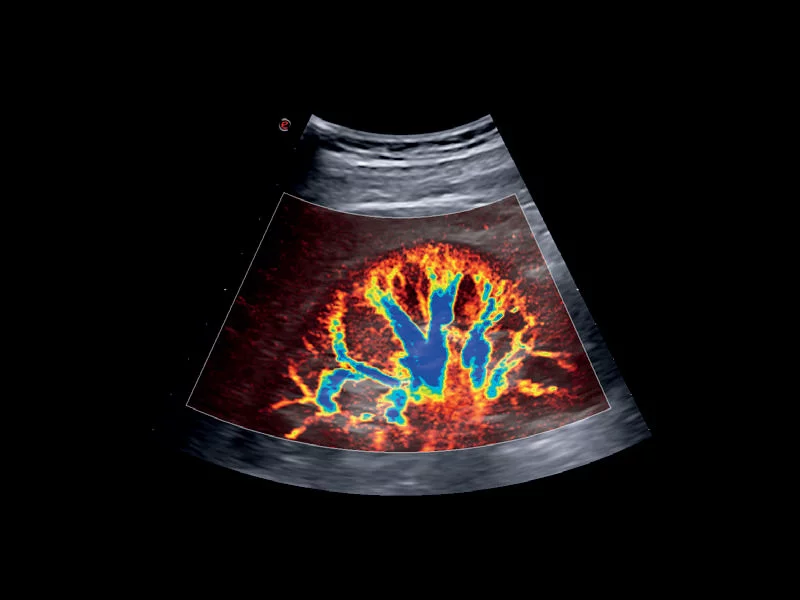
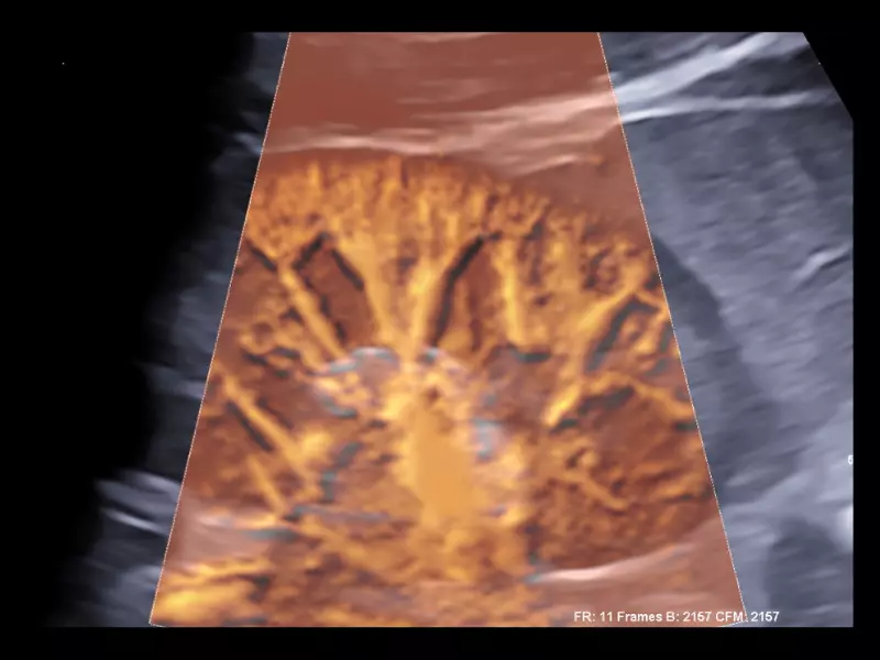
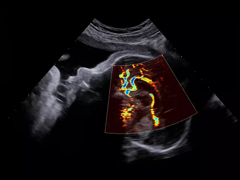
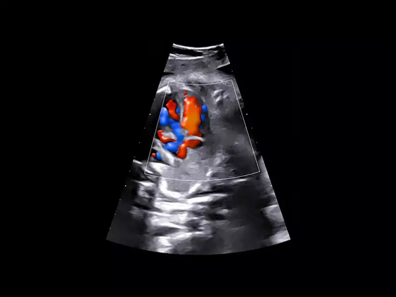
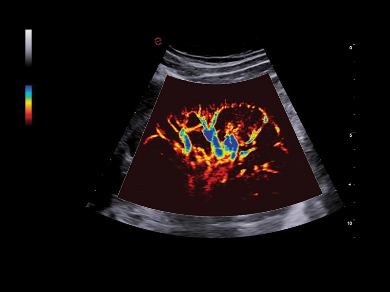
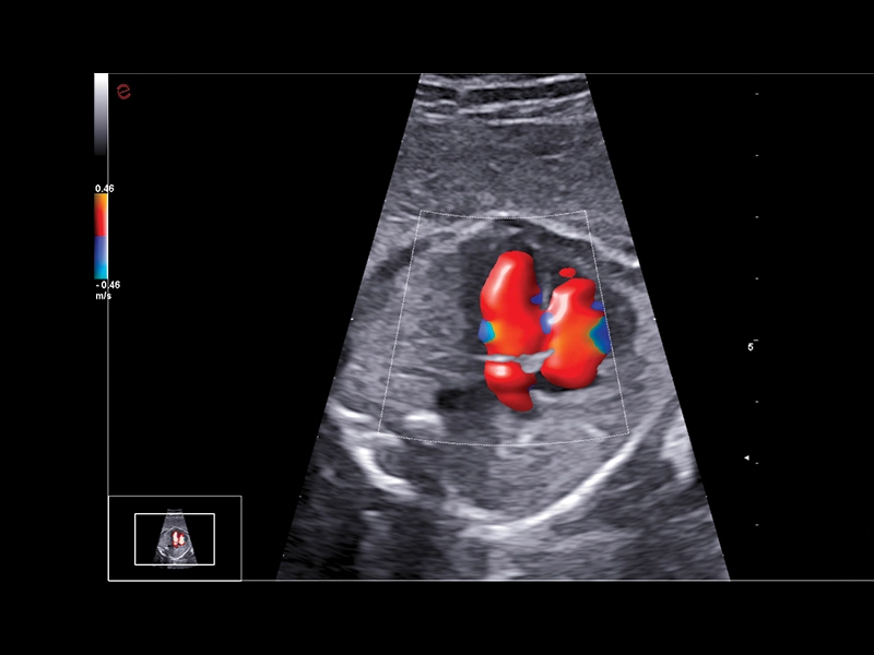
MyLab™Sigma - Fetal Willis circle with Power Doppler
MyLab™Sigma - Fetal Willis circle with Power Doppler
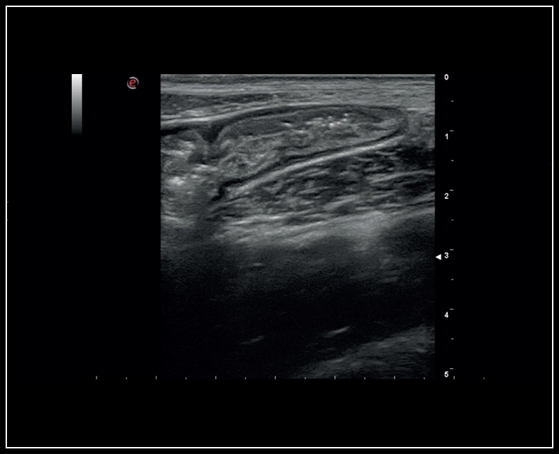
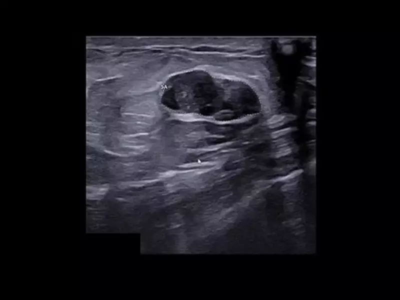
MyLab™Sigma - High Frequency Imaging B-mode of Intestine
MyLab™Sigma - High Frequency Imaging B-mode of Intestine
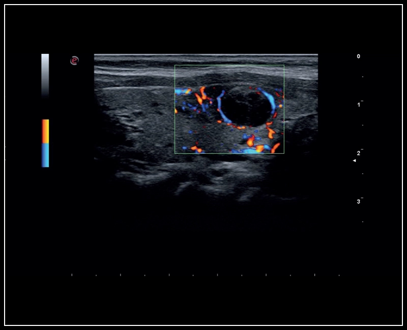
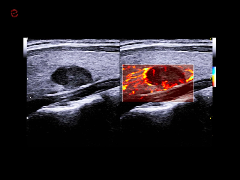
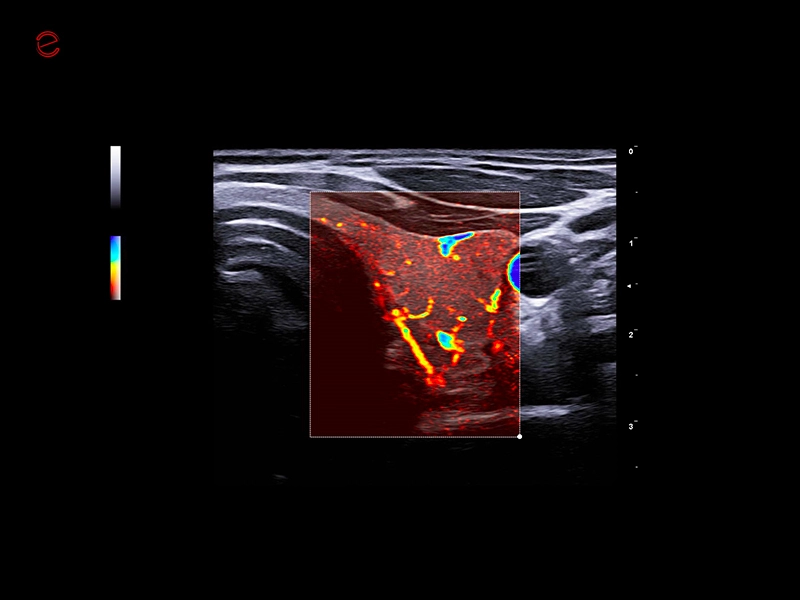
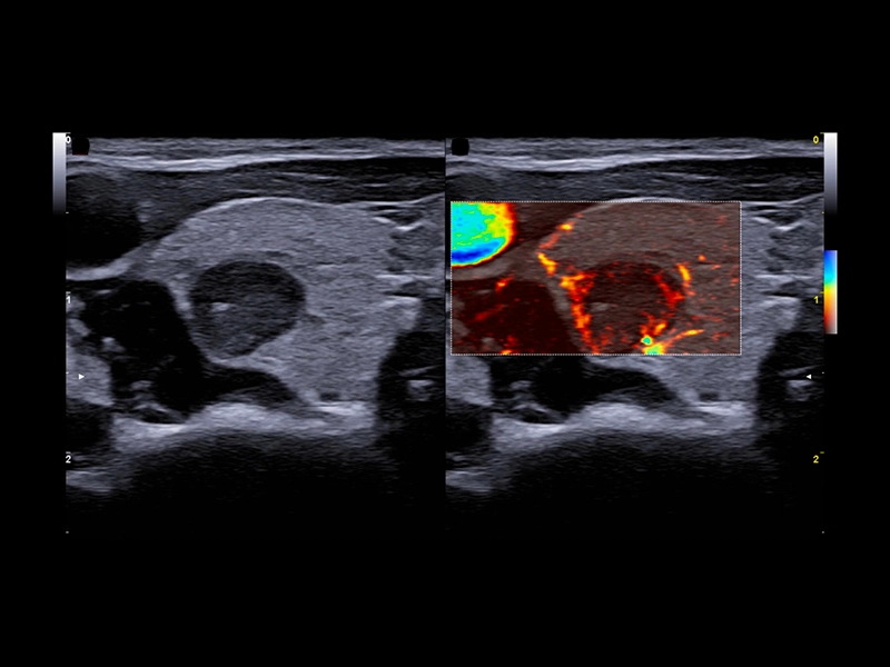
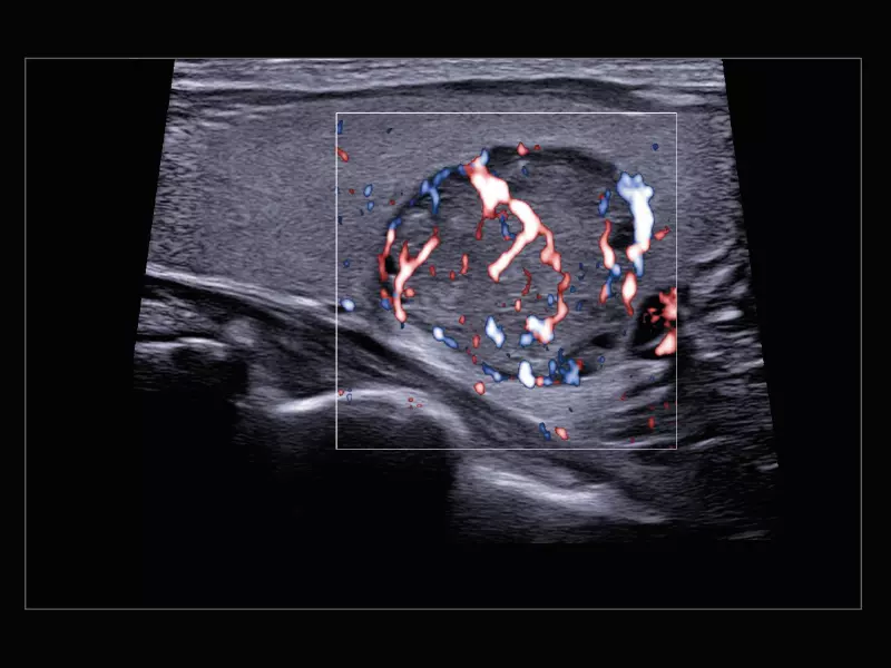
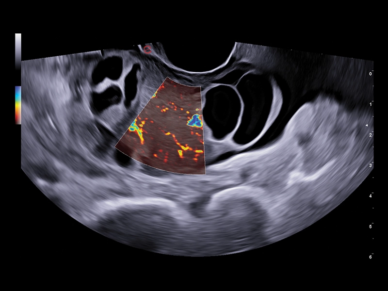
MyLab™Sigma - Thyroid lesion, imaging 2D with Color Doppler
MyLab™Sigma - Thyroid lesion, imaging 2D with Color Doppler
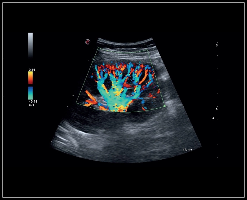
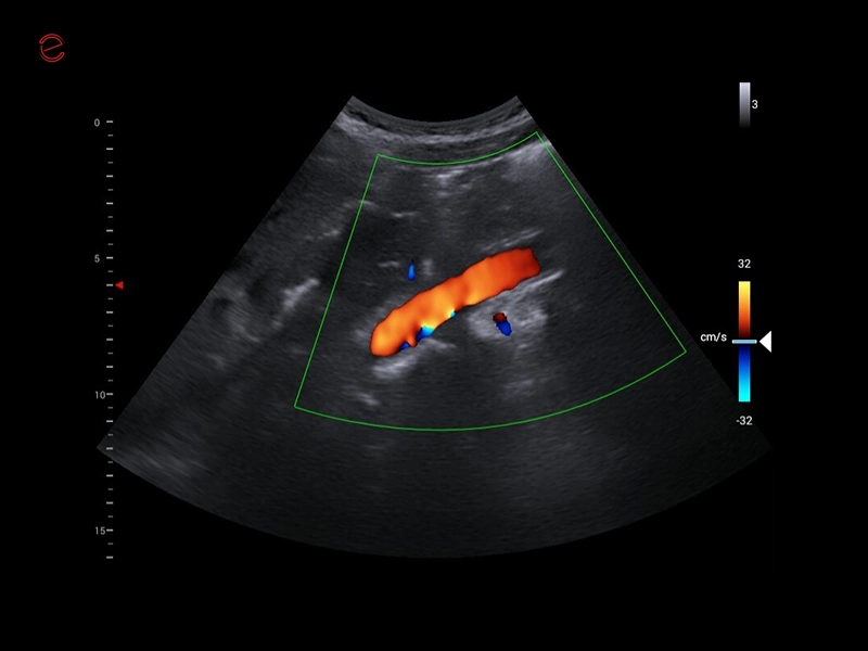
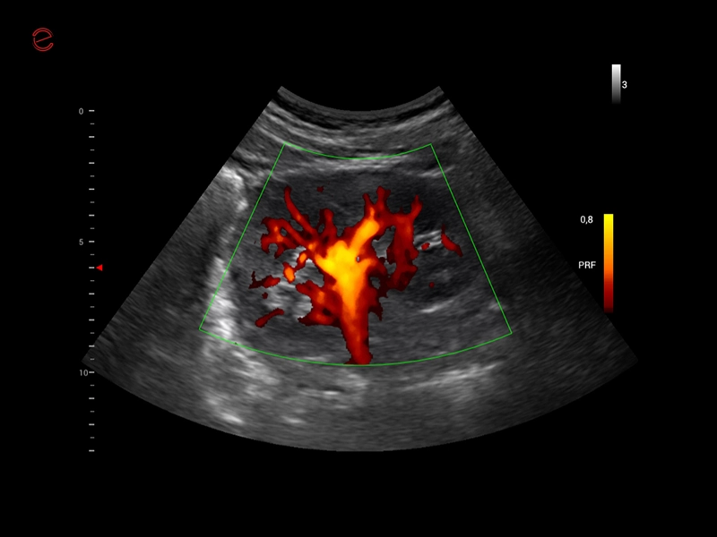
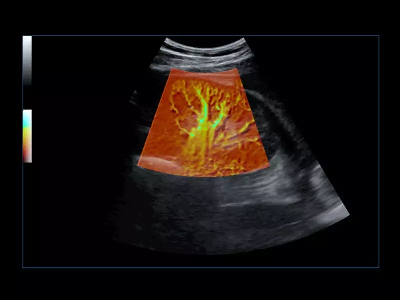
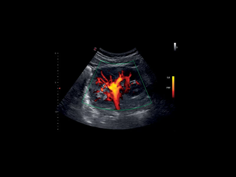
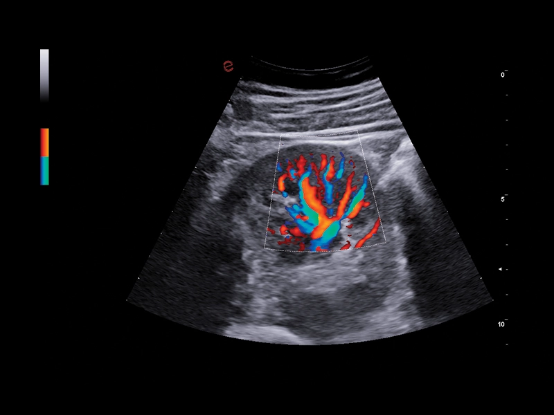
MyLab™Sigma - Kidney perfusion with high sensitivity Color Doppler mode
MyLab™Sigma - Kidney perfusion with high sensitivity Color Doppler mode
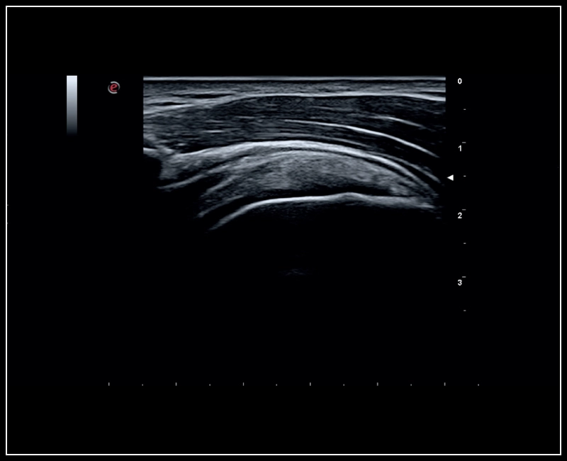
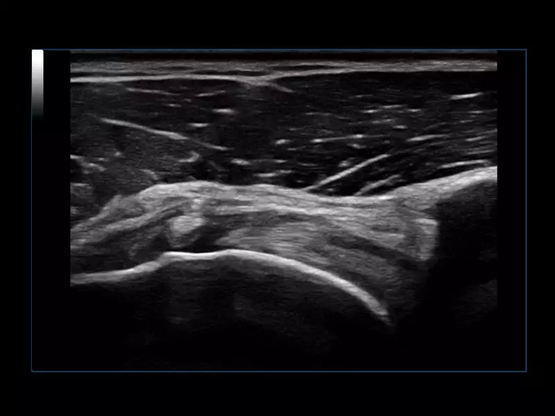
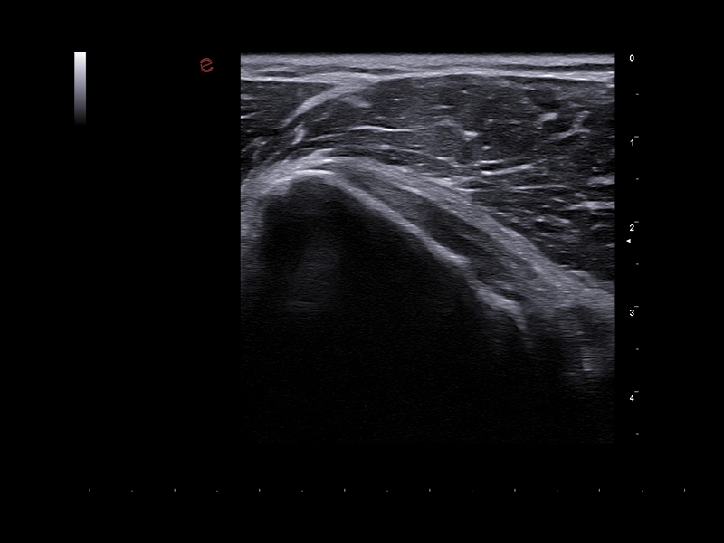
MyLab™Sigma - MSK imaging of the shoulder
MyLab™Sigma - MSK imaging of the shoulder
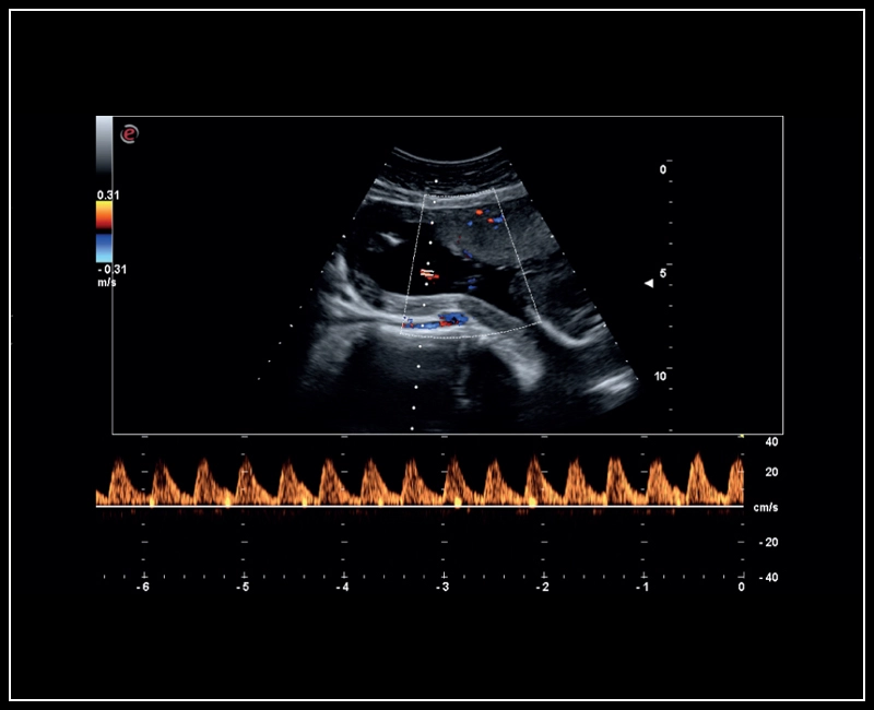
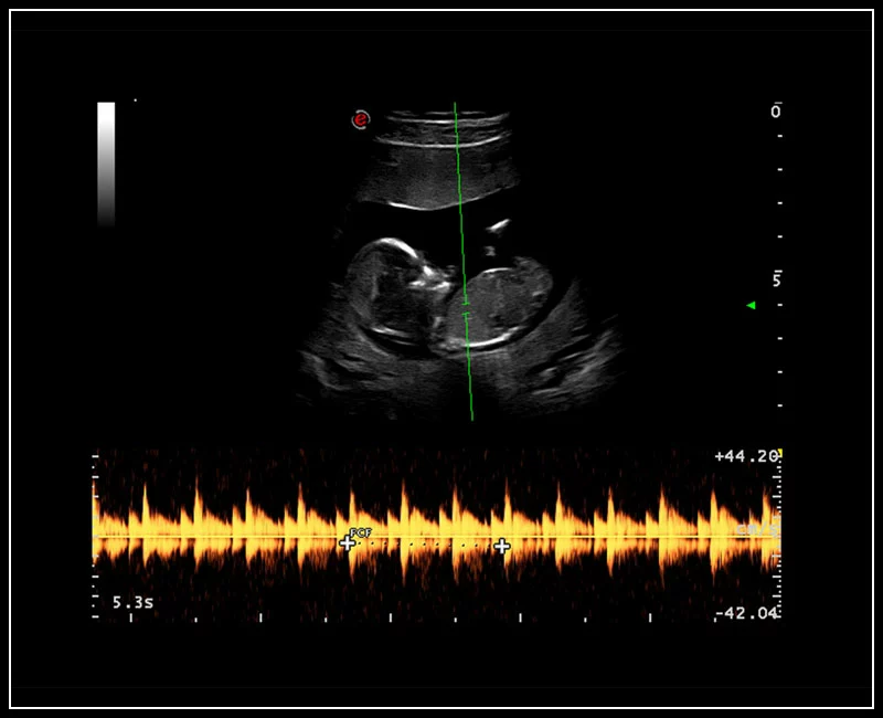
MyLab™Sigma - Umbelical cord PW Doppler mode
MyLab™Sigma - Umbelical cord PW Doppler mode
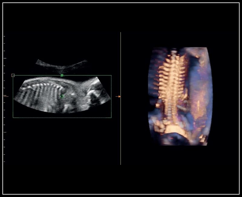
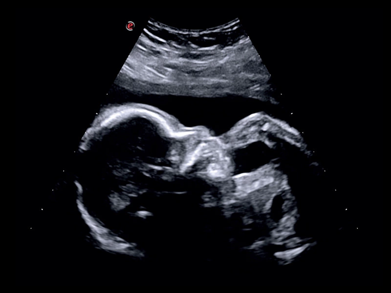
MyLab™Sigma - Semi transparent rendering of fetal spine
MyLab™Sigma - Semi transparent rendering of fetal spine
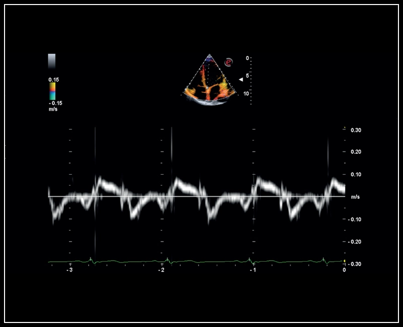
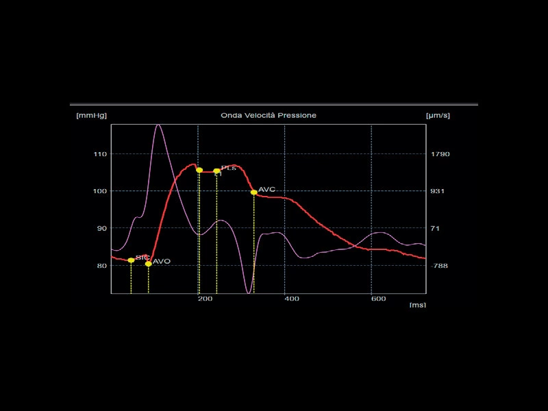
MyLab™Sigma - Mitral valve posterior leaflet analysis with Tissue Velocity Mapping
MyLab™Sigma - Mitral valve posterior leaflet analysis with Tissue Velocity Mapping
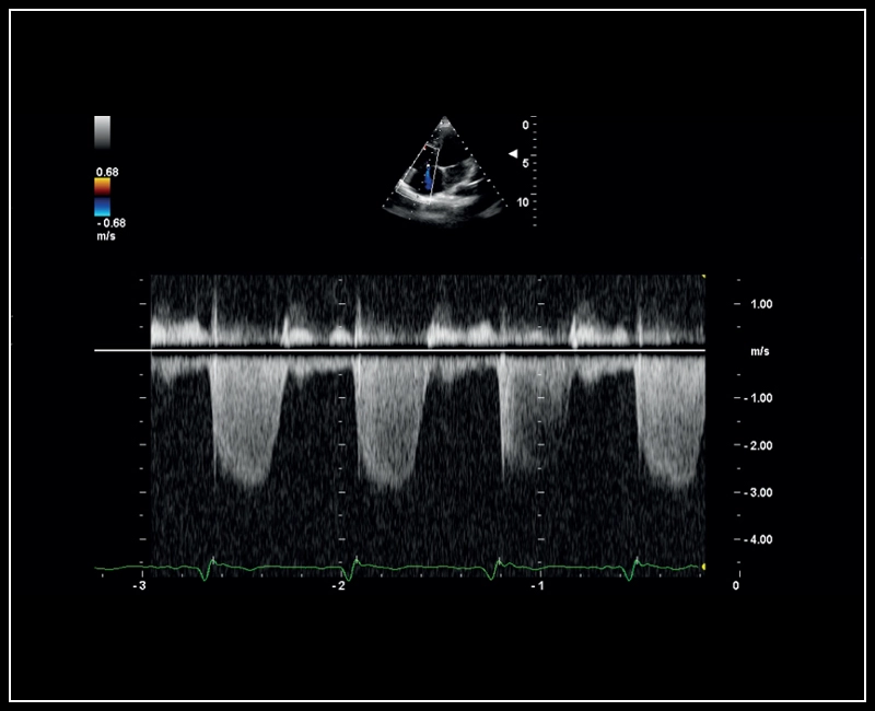
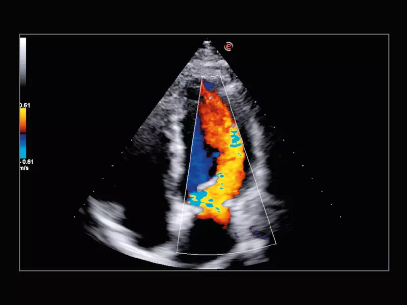
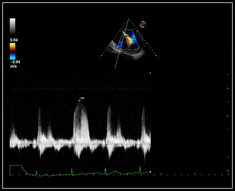
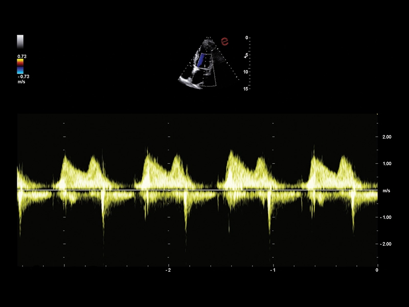
MyLab™Sigma - CW Doppler of Tricuspid regurgitation
MyLab™Sigma - CW Doppler of Tricuspid regurgitation
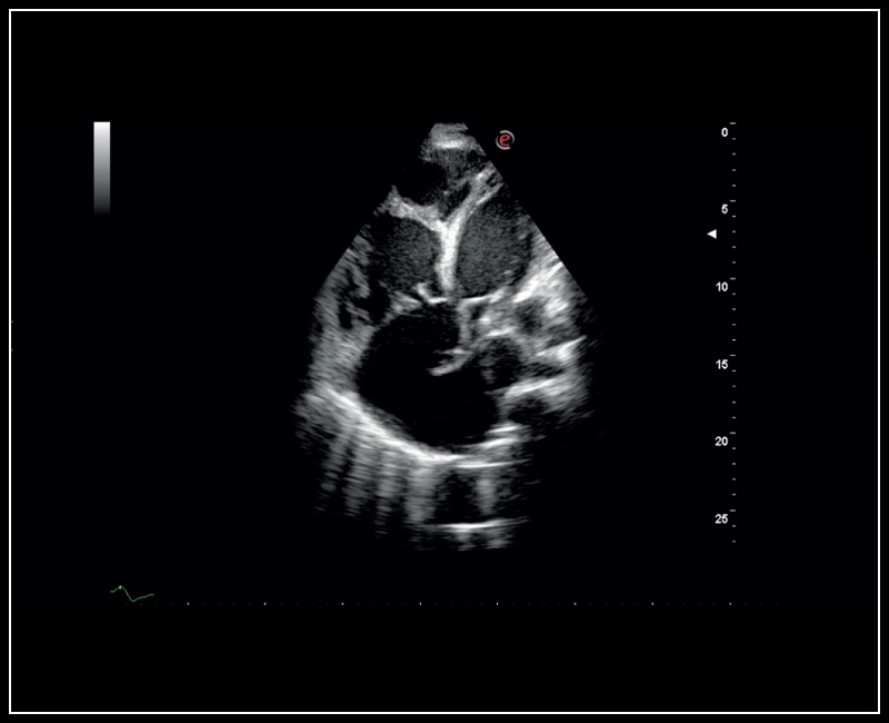
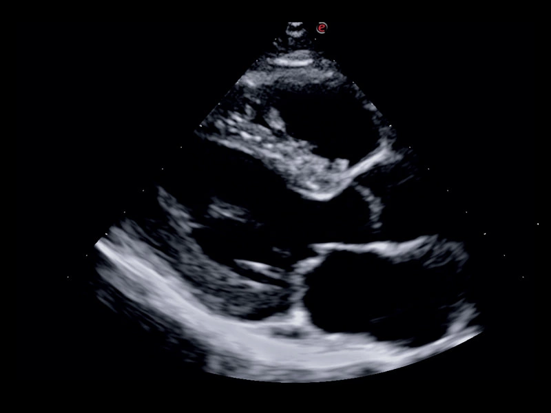
MyLab™Sigma - High definition B-mode cardiac imaging
MyLab™Sigma - High definition B-mode cardiac imaging
MyLab™Sigma - Left Ventricle XStrain 2D analysis
MyLab™Sigma - Left Ventricle XStrain 2D analysis
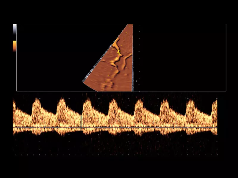
MyLab™Sigma - Mid cerebral artery investigation with PW Doppler mode
MyLab™Sigma - Mid cerebral artery investigation with PW Doppler mode
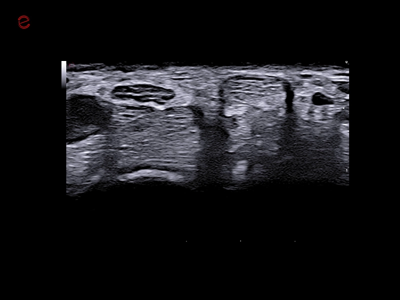
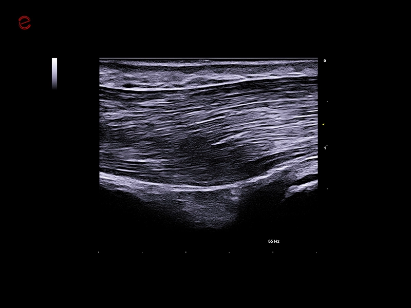
MyLab™Sigma Elite - B-Mode high resolution on median nerve
MyLab™Sigma Elite - B-Mode high resolution on median nerve
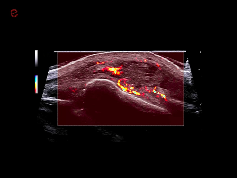
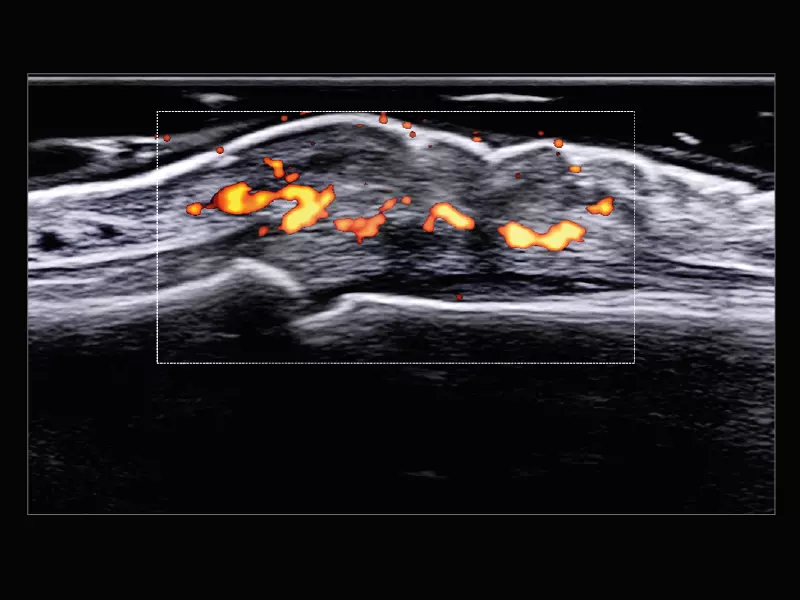
MyLab™Sigma Elite - Power Doppler on acute polyarthritis inflammation area with 18 MHz probe
MyLab™Sigma Elite - Power Doppler on acute polyarthritis inflammation area with 18 MHz probe
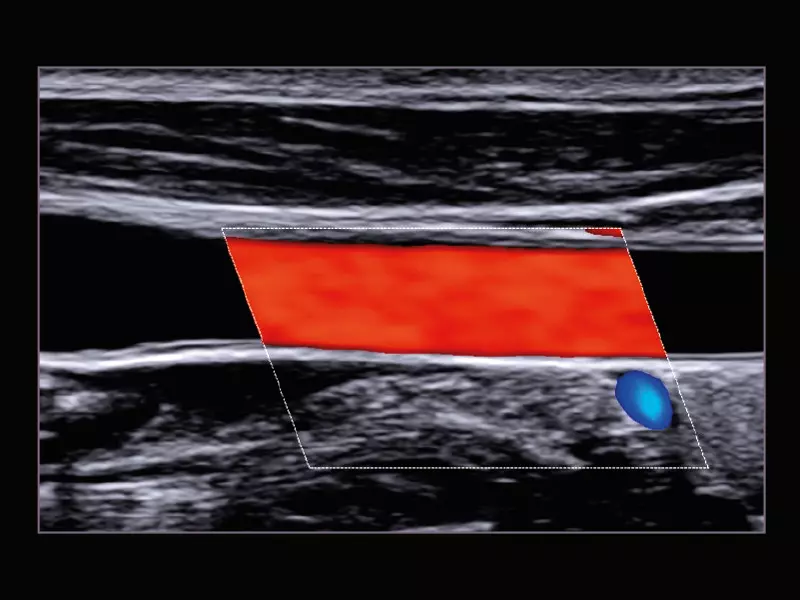
MyLab™Sigma Elite - Temporal Artery exploration with Very High Frequency probe (22 MHz)
MyLab™Sigma Elite - Temporal Artery exploration with Very High Frequency probe (22 MHz)
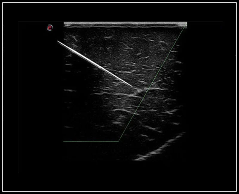
MyLab™Sigma Elite - Needle Enhancement for precise interventional procedures
MyLab™Sigma Elite - Needle Enhancement for precise interventional procedures
MyLab™Sigma Elite - Follow Up in real-time with a second modality
MyLab™Sigma Elite - Follow Up in real-time with a second modality
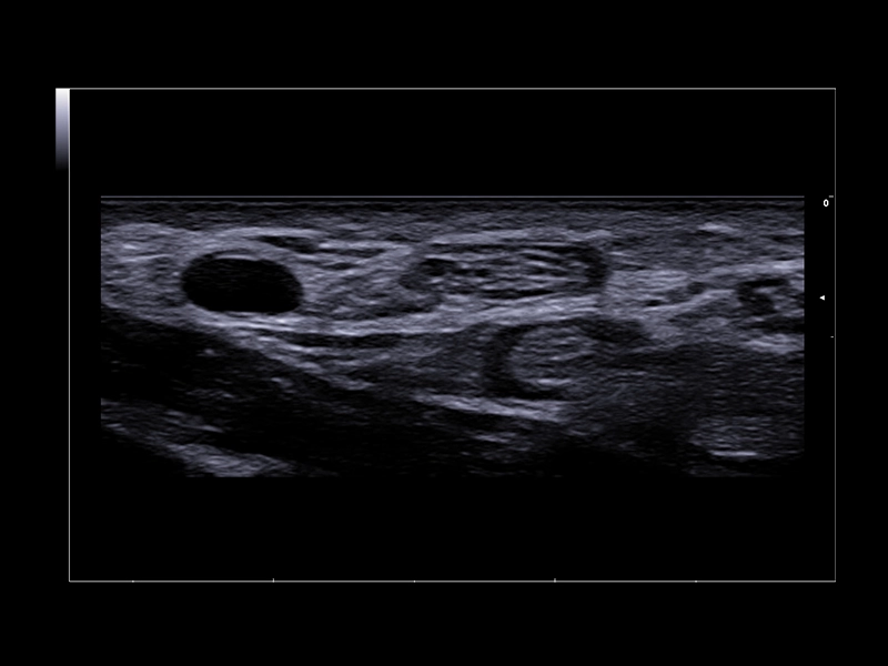
MyLab™X1 - Abdominal liver
MyLab™X1 - Abdominal liver
MyLab™X1 - Apical view CFM
MyLab™X1 - Apical view CFM
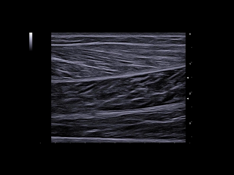
MyLab™X1 - Tendon
MyLab™X1 - Tendon
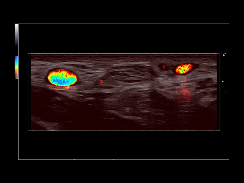
MyLab™X1 - Kidney - PWD
MyLab™X1 - Kidney - PWD
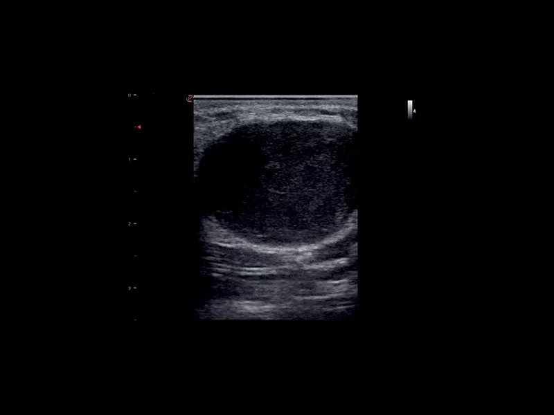
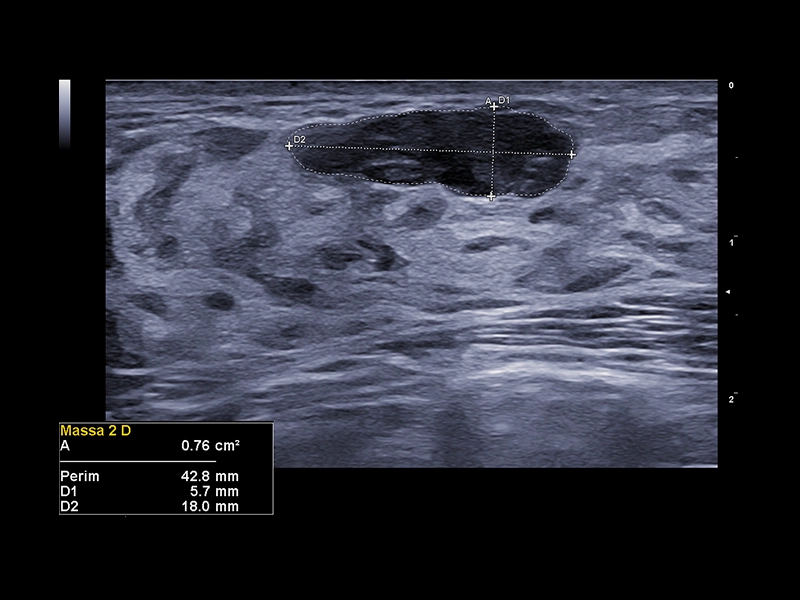
MyLab™X1 - Neck-lump
MyLab™X1 - Neck-lump
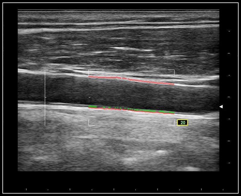
MyLab™X1 - Carotid IMT Calculation
MyLab™X1 - Carotid IMT Calculation
MyLab™X75 - General Imaging 01
MyLab™X75 - General Imaging 01
MyLab™X75 - General Imaging 02
MyLab™X75 - General Imaging 02
MyLab™X75 - General Imaging 03
MyLab™X75 - General Imaging 03
MyLab™X75 - Cardiovascular 04
MyLab™X75 - Cardiovascular 04
MyLab™X75 - General Imaging 04
MyLab™X75 - General Imaging 04
MyLab™X75 - General Imaging 06
MyLab™X75 - General Imaging 06

MyLab™X75 - Cardiovascular 05
MyLab™X75 - Cardiovascular 05
MyLab™X75 - General Imaging 05
MyLab™X75 - General Imaging 05
MyLab™X75 - General Imaging 07
MyLab™X75 - General Imaging 07
MyLab™X75 - Cardiovascular 06
MyLab™X75 - Cardiovascular 06
MyLab™X75 - Cardiovascular 03
MyLab™X75 - Cardiovascular 03
MyLab™X75 - Cardiovascular 02
MyLab™X75 - Cardiovascular 02
MyLab™X75 - Cardiovascular 01
MyLab™X75 - Cardiovascular 01
MyLab™X75 - Women's Health 05
MyLab™X75 - Women's Health 05
MyLab™X75 - Women's Health 04
MyLab™X75 - Women's Health 04
MyLab™X75 - Women's Health 03
MyLab™X75 - Women's Health 03
MyLab™X75 - Women's Health 02
MyLab™X75 - Women's Health 02
MyLab™X75 - Women's Health 01
MyLab™X75 - Women's Health 01
MyLab™X8 Platform - Very high frequency exploration of up to 24MHz
MyLab™X8 Platform - Very high frequency exploration of up to 24MHz
MyLab™X8 Platform - Lesion with ElaXto
MyLab™X8 Platform - Lesion with ElaXto
MyLab™X8 Platform - XView algorithm for increased visibility
MyLab™X8 Platform - XView algorithm for increased visibility
MyLab™X8 Platform - CPI with color Doppler
MyLab™X8 Platform - CPI with color Doppler
MyLab™X8 Platform - Real-time measurement of the Intima Media with QIMT
MyLab™X8 Platform - Real-time measurement of the Intima Media with QIMT
MyLab™X8 Platform - Zero-click EF measurement with Uterus - XLight 3D/4D exploration AutoEF
MyLab™X8 Platform - Zero-click EF measurement with Uterus - XLight 3D/4D exploration AutoEF
MyLab™X8 Platform - Zero-click strain evaluation with XStrain™2D
MyLab™X8 Platform - Zero-click strain evaluation with XStrain™2D
MyLab™X8 Platform - XLight 3D/4D exploration
MyLab™X8 Platform - XLight 3D/4D exploration
MyLab™X8 Platform - Tissue perfusion visualisation with microV
MyLab™X8 Platform - Tissue perfusion visualisation with microV
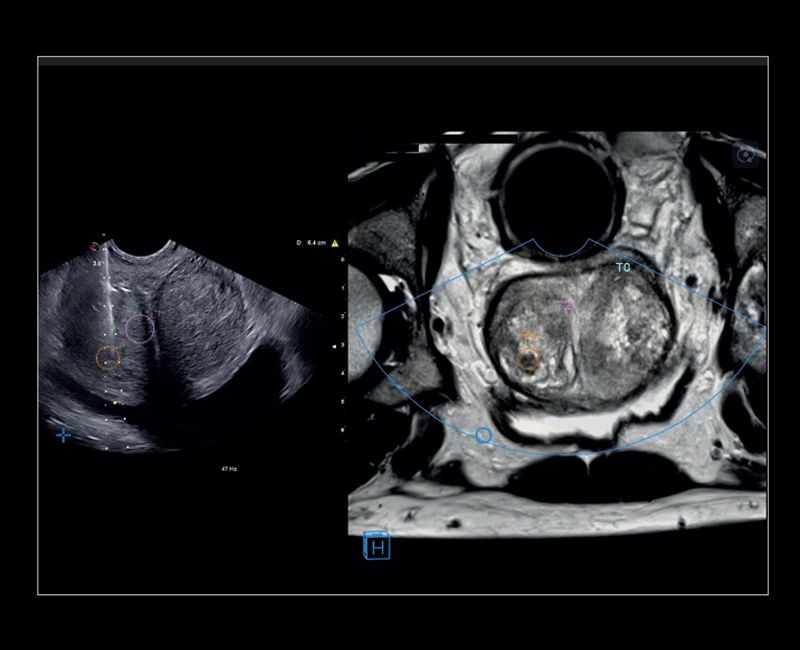
MyLab™X8 Platform - Prostate biopsy with Virtual Navigator fusion imaging 3D
MyLab™X8 Platform - Prostate biopsy with Virtual Navigator fusion imaging 3D
MyLab™X8 Platform - Volumetric model of the left function with XStrain4D
MyLab™X8 Platform - Volumetric model of the left function with XStrain4D
MyLab™X8 Platform - Evolution of the inflammation with Follow Up and Q-Pack
MyLab™X8 Platform - Evolution of the inflammation with Follow Up and Q-Pack
MyLab™X8 Platform - Contrast session with CnTI™ and Q-Pack
MyLab™X8 Platform - Contrast session with CnTI™ and Q-Pack
MyLab™X8 Platform - Stiffness quantification with Q-ElaXto
MyLab™X8 Platform - Stiffness quantification with Q-ElaXto
MyLab™X8 Platform - Accurate multimodality diagnosis with Follow Up and BodyMap
MyLab™X8 Platform - Accurate multimodality diagnosis with Follow Up and BodyMap
MyLab™X9 - General Imaging 01
MyLab™X9 - General Imaging 01
MyLab™X9 - General Imaging 02
MyLab™X9 - General Imaging 02
MyLab™X9 - General Imaging 03
MyLab™X9 - General Imaging 03
MyLab™X9 - General Imaging 04
MyLab™X9 - General Imaging 04
MyLab™X9 - General Imaging 05
MyLab™X9 - General Imaging 05
MyLab™X9 - General Imaging 06
MyLab™X9 - General Imaging 06
MyLab™X9 - Liver 01
MyLab™X9 - Liver 01
MyLab™X9 - Liver 02
MyLab™X9 - Liver 02
MyLab™X9 - Liver 03
MyLab™X9 - Liver 03
MyLab™X9 - Liver 04
MyLab™X9 - Liver 04
MyLab™X9 - Liver 05
MyLab™X9 - Liver 05
MyLab™X9 - Interventional
MyLab™X9 - Interventional
MyLab™X9 - Urology 01
MyLab™X9 - Urology 01
MyLab™X9 - Urology 02
MyLab™X9 - Urology 02
MyLab™X9 - Urology 03
MyLab™X9 - Urology 03
MyLab™X9 - Urology 04
MyLab™X9 - Urology 04
MyLab™X9 - Urology 05
MyLab™X9 - Urology 05
MyLab™X9 - Urology 06
MyLab™X9 - Urology 06
MyLab™X9 - Breast 01
MyLab™X9 - Breast 01
MyLab™X9 - Breast 02
MyLab™X9 - Breast 02
MyLab™X9 - Breast 03
MyLab™X9 - Breast 03
MyLab™X9 - Breast 04
MyLab™X9 - Breast 04
MyLab™X9 - Breast 05
MyLab™X9 - Breast 05
MyLab™X9 - Breast 06
MyLab™X9 - Breast 06
MyLab™X9 - Superficial 01
MyLab™X9 - Superficial 01
MyLab™X9 - Superficial 02
MyLab™X9 - Superficial 02
MyLab™X9 - Superficial 03
MyLab™X9 - Superficial 03
MyLab™X9 - Superficial 04
MyLab™X9 - Superficial 04
MyLab™X9 - Shared Service 01
MyLab™X9 - Shared Service 01
MyLab™X9 - Shared Service 02
MyLab™X9 - Shared Service 02
MyLab™X9 - Shared Service 03
MyLab™X9 - Shared Service 03
MyLab™X9 - Shared Service 04
MyLab™X9 - Shared Service 04
MyLab™X9 - Shared Service 05
MyLab™X9 - Shared Service 05
MyLab™X9 - Shared Service 06
MyLab™X9 - Shared Service 06
MyLab™X9 - Shared Service 07
MyLab™X9 - Shared Service 07
MyLab™X9 - Shared Service 08
MyLab™X9 - Shared Service 08
MyLab™X9 - Shared Service 09
MyLab™X9 - Shared Service 09
MyLab™X9 - Shared Service 10
MyLab™X9 - Shared Service 10
MyLab™X9 - Shared Service 11
MyLab™X9 - Shared Service 11
MyLab™X9 - Shared Service 12
MyLab™X9 - Shared Service 12
MyLab™X90 - Cardiovascular 01
MyLab™X90 - Cardiovascular 01
MyLab™X90 - General Imaging 01
MyLab™X90 - General Imaging 01
MyLab™X90 - Women's Health 01
MyLab™X90 - Women's Health 01
MyLab™X90 - Cardiovascular 02
MyLab™X90 - Cardiovascular 02
MyLab™X90 - Cardiovascular 03
MyLab™X90 - Cardiovascular 03
MyLab™X90 - Cardiovascular 04
MyLab™X90 - Cardiovascular 04
MyLab™X90 - Cardiovascular 05
MyLab™X90 - Cardiovascular 05
MyLab™X90 - Urology 01
MyLab™X90 - Urology 01
MyLab™X90 - Urology 02
MyLab™X90 - Urology 02
MyLab™X90 - Urology 03
MyLab™X90 - Urology 03
MyLab™X90 - Urology 04
MyLab™X90 - Urology 04
MyLab™X90 - Women's Health 02
MyLab™X90 - Women's Health 02
MyLab™X90 - General Imaging 02
MyLab™X90 - General Imaging 02
MyLab™X90 - MSK 01
MyLab™X90 - MSK 01
MyLab™X90 - MSK 02
MyLab™X90 - MSK 02
MyLab™X90 - General Imaging 02
MyLab™X90 - General Imaging 02
MyLab™X90 - Liver 01
MyLab™X90 - Liver 01
MyLab™X90 - Liver 02
MyLab™X90 - Liver 02
MyLab™X90 - Liver 03
MyLab™X90 - Liver 03
MyLab™X90 - General Imaging 03
MyLab™X90 - General Imaging 03
MyLab™X90 - Cardiovascular 06
MyLab™X90 - Cardiovascular 06
MyLab™X90 - Women's Health 03
MyLab™X90 - Women's Health 03
MyLab™X90 - Women's Health 04
MyLab™X90 - Women's Health 04
MyLab™X90 - General Imaging 04
MyLab™X90 - General Imaging 04
MyLab™X90 - General Imaging 05
MyLab™X90 - General Imaging 05
MyLab™X90 - Automatic Plan recognition Automatic biometric plan recognition for fetal measurements
MyLab™X90 - Automatic Plan recognition Automatic biometric plan recognition for fetal measurements
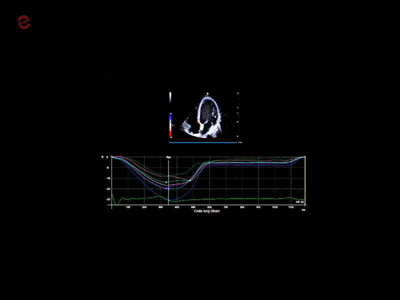
MyLab™X90 - XStrain™ LV Automatic assessment of global longitudinal strain in the left ventricle
MyLab™X90 - XStrain™ LV Automatic assessment of global longitudinal strain in the left ventricle
MyLab™X90 - BreastNav™ MRI Automatic segmentation of the breast MRI and real-time fusion based on an adaptive 3D mod
MyLab™X90 - BreastNav™ MRI Automatic segmentation of the breast MRI and real-time fusion based on an adaptive 3D mod
MyLab™X90 - Women's Health 04
MyLab™X90 - Women's Health 04
MyLab™X90 - Cardiovascular 07
MyLab™X90 - Cardiovascular 07
MyLab™X90 - AutoEF Automatic Ejection fraction assessment of the left ventricle
MyLab™X90 - AutoEF Automatic Ejection fraction assessment of the left ventricle
MyLab™X90 - Women's Health 05
MyLab™X90 - Women's Health 05
MyLab™X90 - Women's Health 06
MyLab™X90 - Women's Health 06
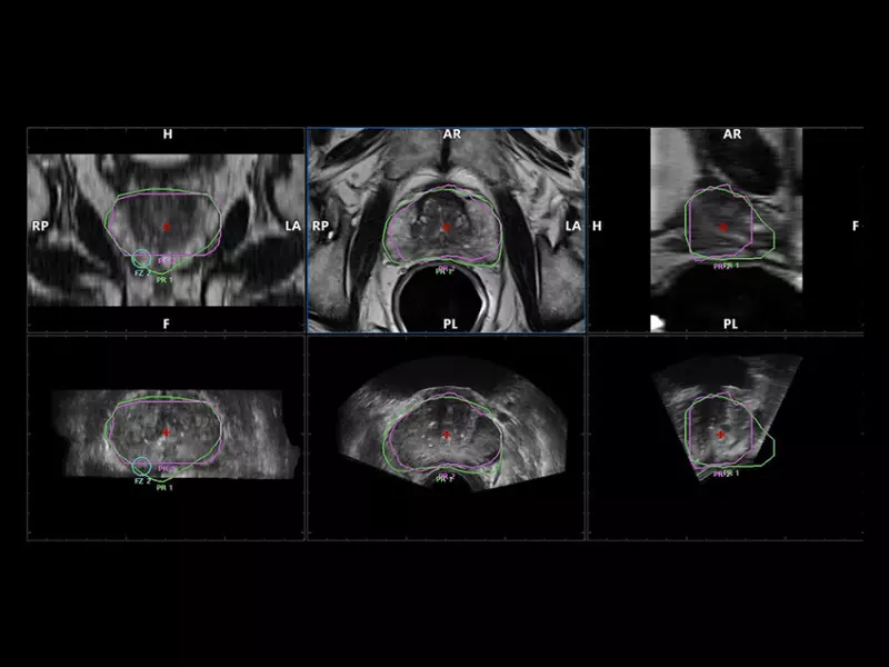
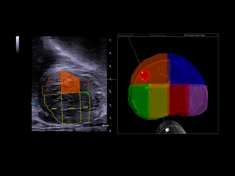
MyLab™X90 - UroFusion Automatic segmentation of the prostate MRI/US, followed by autoregistration of both modalities for targeted biopsies
MyLab™X90 - UroFusion Automatic segmentation of the prostate MRI/US, followed by autoregistration of both modalities for targeted biopsies
MyLab™X90 - BMA: Automatic proposal of breast lesion classification, inside a region of clinical interest (ROI)
MyLab™X90 - BMA: Automatic proposal of breast lesion classification, inside a region of clinical interest (ROI)
MyLab™X90 - eDetect Automatic contouring of thyroid and breast lesions in suspicious areas suggested (ROI) by the physicians
MyLab™X90 - eDetect Automatic contouring of thyroid and breast lesions in suspicious areas suggested (ROI) by the physicians
MyLab™X90 - Liver 04
MyLab™X90 - Liver 04
MyLab™X90 - AutoOB Automatic proposals for fetal biometric measurements
MyLab™X90 - AutoOB Automatic proposals for fetal biometric measurements
MyLab™X90 - General Imaging 06
MyLab™X90 - General Imaging 06
MyLab™X90 - General Imaging 07
MyLab™X90 - General Imaging 07
MyLab™X90 - General Imaging 08
MyLab™X90 - General Imaging 08
MyLab™X90 - Women's Health 07
MyLab™X90 - Women's Health 07

MyLab™X5 - AutoEF
MyLab™X5 - AutoEF
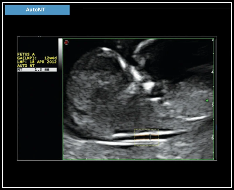
MyLab™X5 - AutoNT
MyLab™X5 - AutoNT
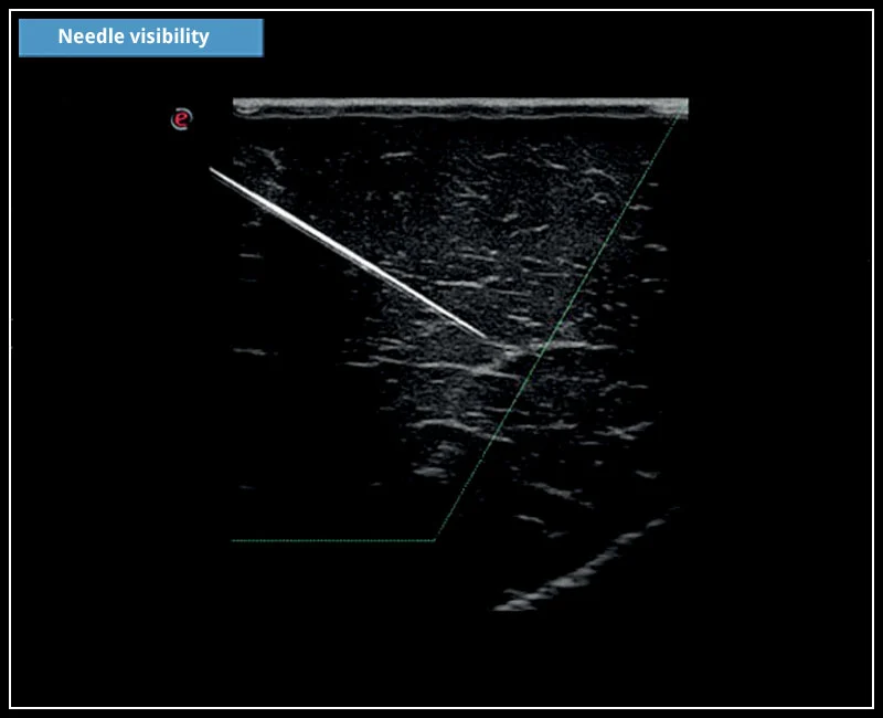
MyLab™X5 - Needle visibility
MyLab™X5 - Needle visibility
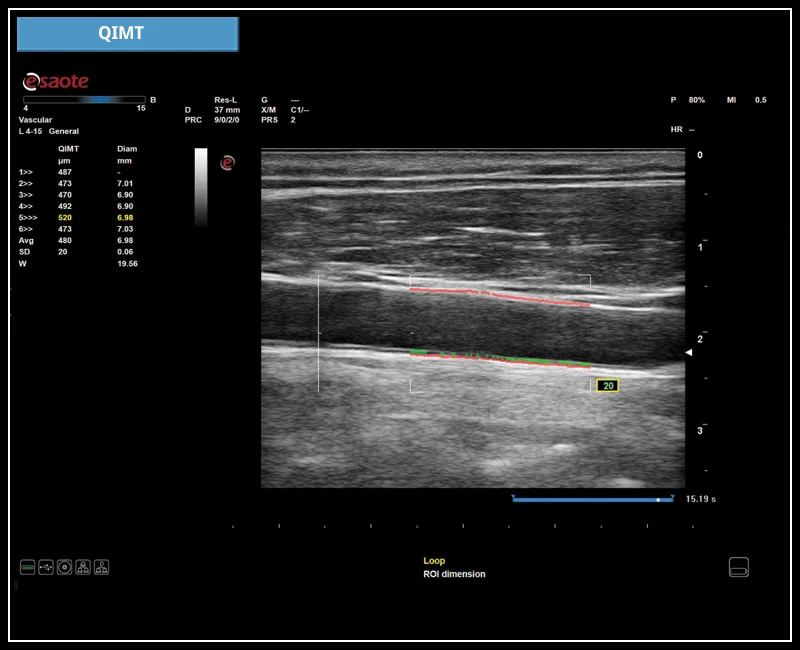
MyLab™X5 - QIMT
MyLab™X5 - QIMT
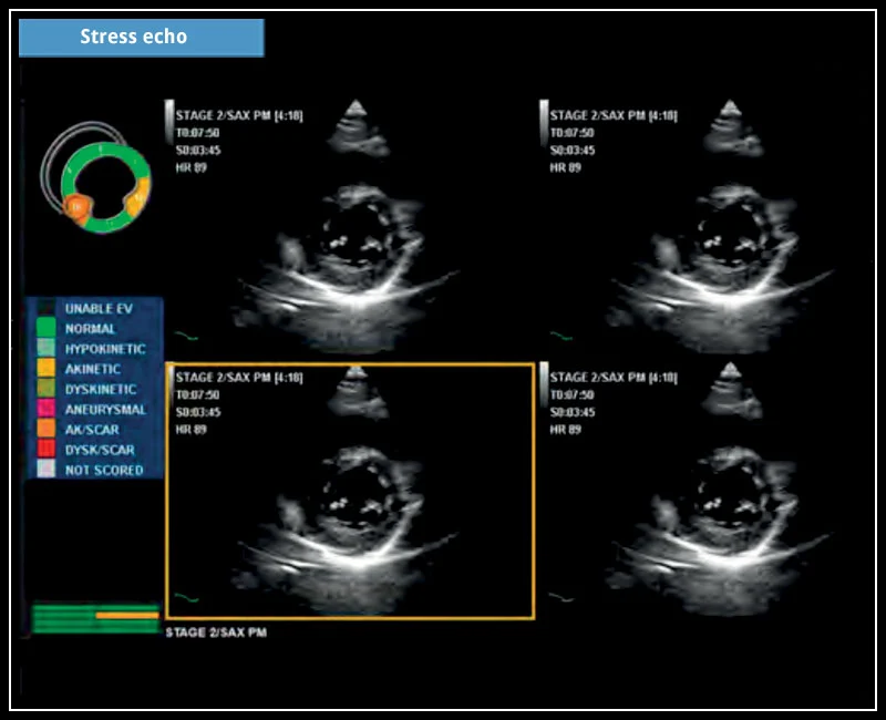
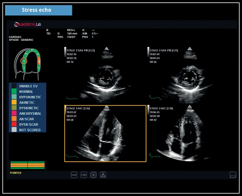
MyLab™X5 - Stress echo
MyLab™X5 - Stress echo

MyLab™X5 - XStrain
MyLab™X5 - XStrain

MyLab™X6 - AutoEF
MyLab™X6 - AutoEF
MyLab™X6 - AutoNT
MyLab™X6 - AutoNT
MyLab™X6 - ElaXto
MyLab™X6 - ElaXto

MyLab™X6 - Needle Visibility
MyLab™X6 - Needle Visibility
MyLab™X6 - QIMT
MyLab™X6 - QIMT
MyLab™X6 - Stress echo
MyLab™X6 - Stress echo

MyLab™X6 - XLight
MyLab™X6 - XLight

MyLab™X6 - XStrain
MyLab™X6 - XStrain
MyLab™X7 - AutoEF
MyLab™X7 - AutoEF

MyLab™X7 - AutoNT
MyLab™X7 - AutoNT
MyLab™X7 - CnTI™
MyLab™X7 - CnTI™
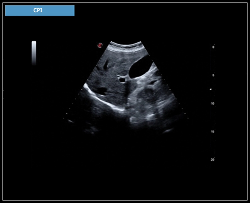
MyLab™X7 - CPI
MyLab™X7 - CPI

MyLab™X7 - ElaXto
MyLab™X7 - ElaXto

MyLab™X7 - microV
MyLab™X7 - microV
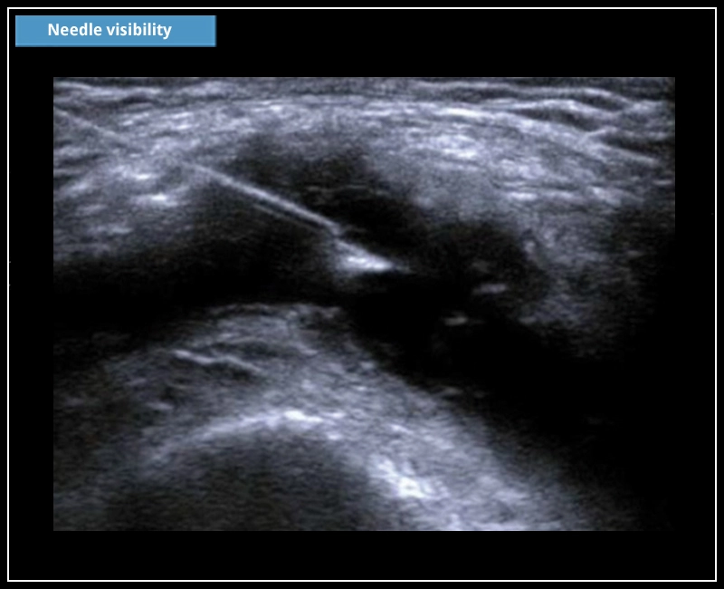
MyLab™X7 - Needle visibility
MyLab™X7 - Needle visibility
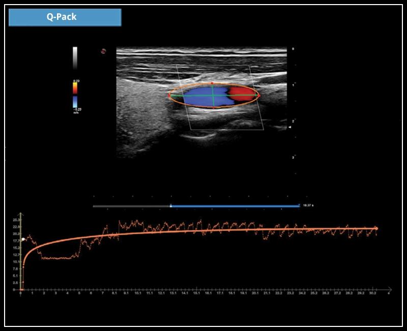
MyLab™X7 - Q-Pack
MyLab™X7 - Q-Pack
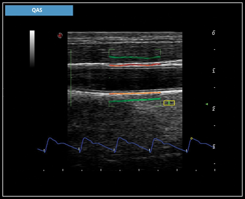
MyLab™X7 - QAS
MyLab™X7 - QAS
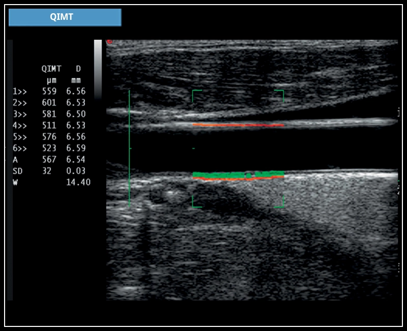
MyLab™X7 - QIMT
MyLab™X7 - QIMT
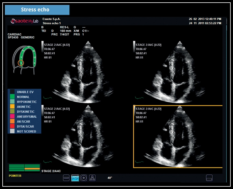
MyLab™X7 - Stress echo
MyLab™X7 - Stress echo
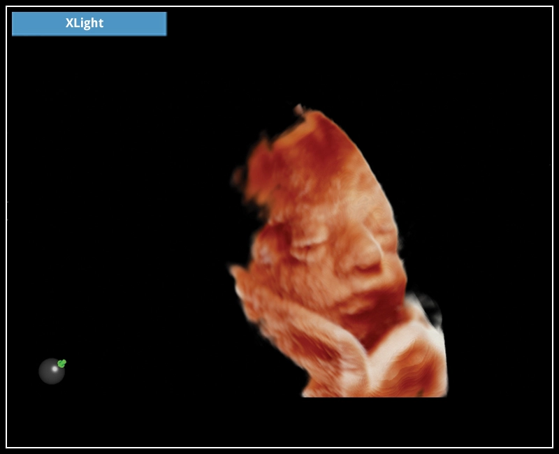
MyLab™X7 - XLight
MyLab™X7 - XLight
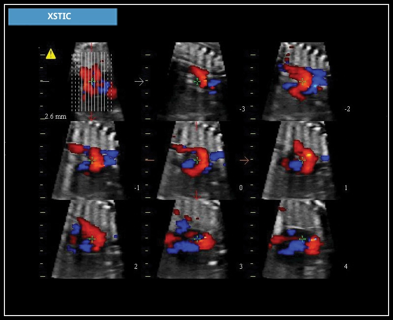
MyLab™X7 - XSTIC
MyLab™X7 - XSTIC
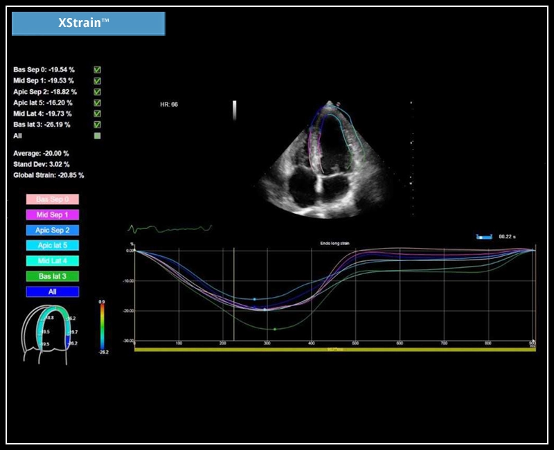
MyLab™X7 - XStrain
MyLab™X7 - XStrain
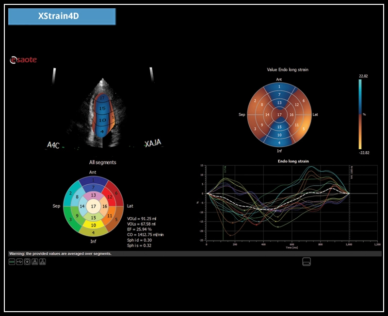
MyLab™X7 - XStrain4D
MyLab™X7 - XStrain4D
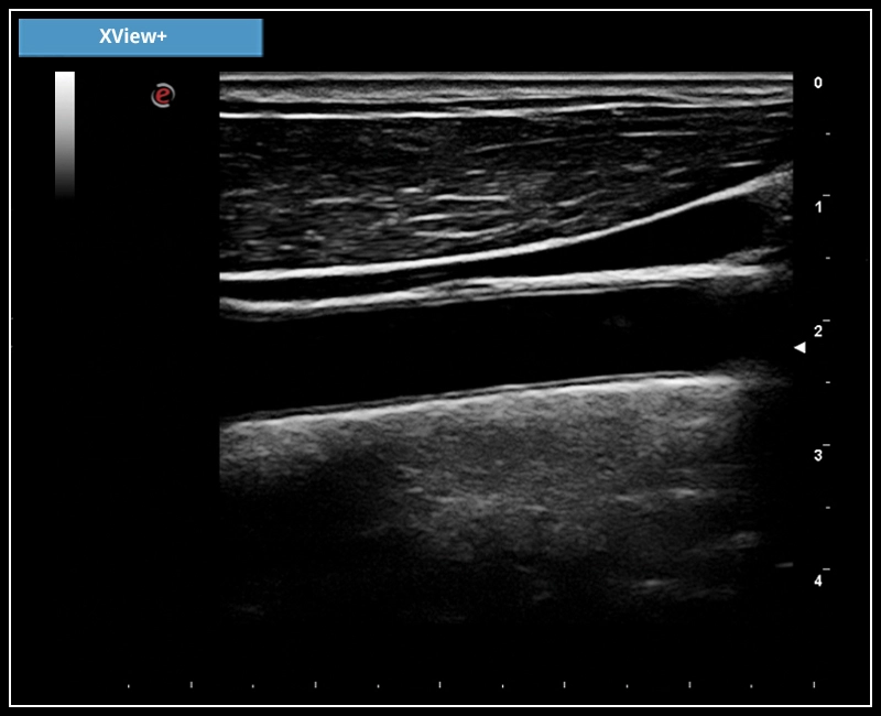
MyLab™X7 - XView+
MyLab™X7 - XView+
MyLab™Omega - AutoNT
MyLab™Omega - AutoNT
MyLab™Omega - BabyFace using XLight
MyLab™Omega - BabyFace using XLight
MyLab™Omega - Common Carotid Artery CFM
MyLab™Omega - Common Carotid Artery CFM
MyLab™Omega - CPI and XView liver cyst visualization
MyLab™Omega - CPI and XView liver cyst visualization
MyLab™Omega - CW Doppler
MyLab™Omega - CW Doppler
MyLab™Omega - Dual ElaXto on breast cyst
MyLab™Omega - Dual ElaXto on breast cyst
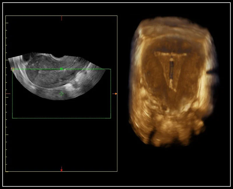
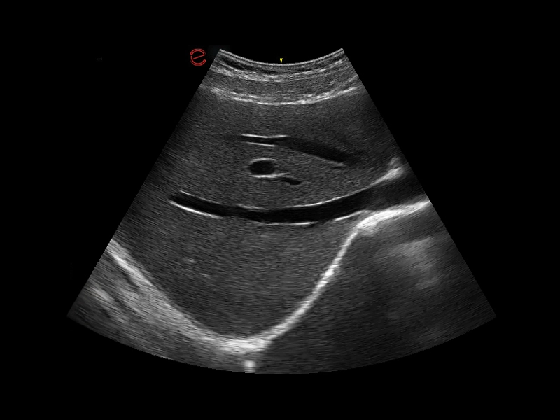
MyLab™Omega - IUD visualization using 3D endocavitary probe
MyLab™Omega - IUD visualization using 3D endocavitary probe
MyLab™Omega - Liver metastasis using CnTI™
MyLab™Omega - Liver metastasis using CnTI™
MyLab™Omega - Optima needle tip visualization using Needle Enhancement
MyLab™Omega - Optima needle tip visualization using Needle Enhancement
MyLab™Omega - RF QIMT
MyLab™Omega - RF QIMT
MyLab™Omega - Short axis big vessel right ventricle CFM
MyLab™Omega - Short axis big vessel right ventricle CFM
MyLab™Omega - Stress Echo
MyLab™Omega - Stress Echo
MyLab™Omega - Transcranial doppler
MyLab™Omega - Transcranial doppler
MyLab™Omega - Zero-Click AutoEF
MyLab™Omega - Zero-Click AutoEF
MyLab™A50 - AutoEF
MyLab™A50 - AutoEF
MyLab™A50 - autoOB
MyLab™A50 - autoOB
MyLab™A50 - CFM liver
MyLab™A50 - CFM liver
MyLab™A50 - ElaXto
MyLab™A50 - ElaXto
MyLab™A50 - Face profile
MyLab™A50 - Face profile

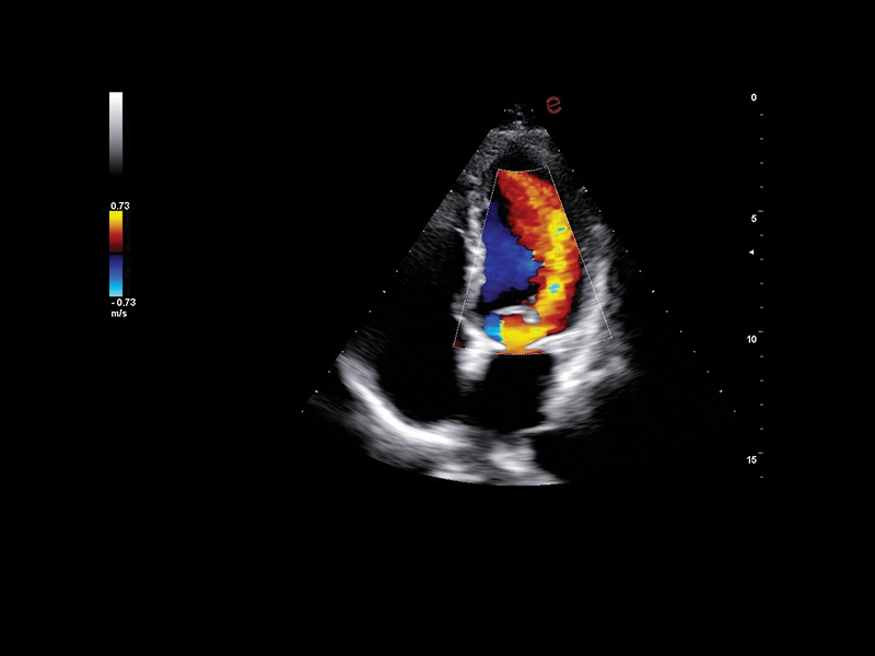
MyLab™A50 - Heart CFM
MyLab™A50 - Heart CFM
MyLab™A50 - Heart CW
MyLab™A50 - Heart CW
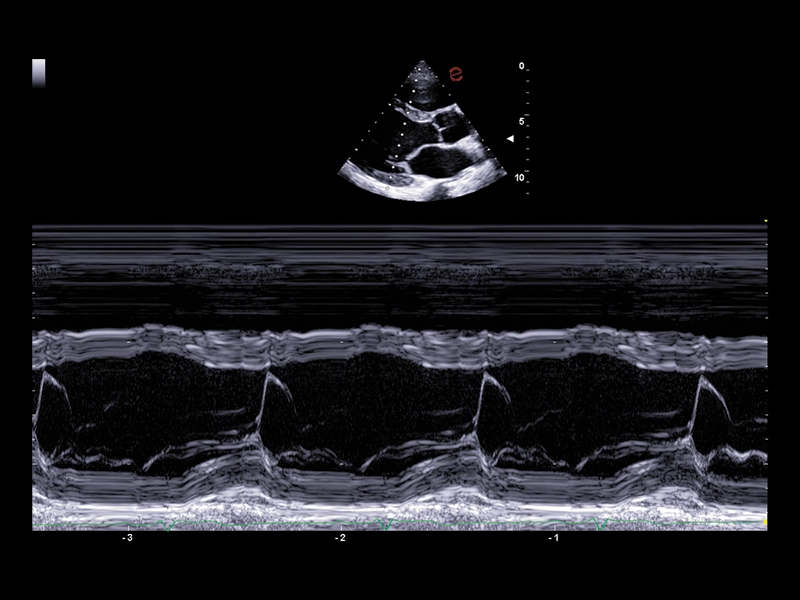
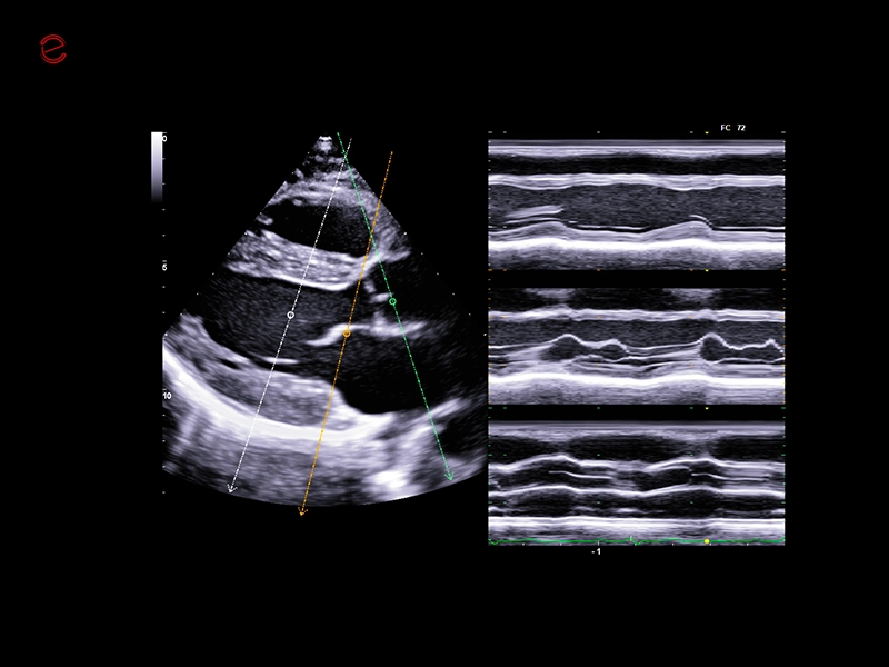
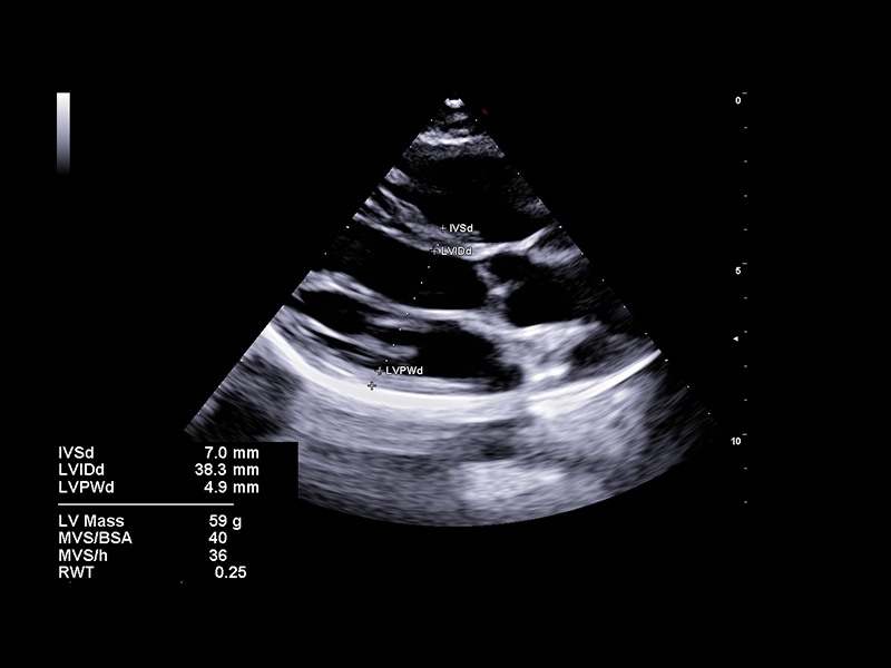
MyLab™A50 - Heart Mmode
MyLab™A50 - Heart Mmode
MyLab™A50 - HY microV
MyLab™A50 - HY microV
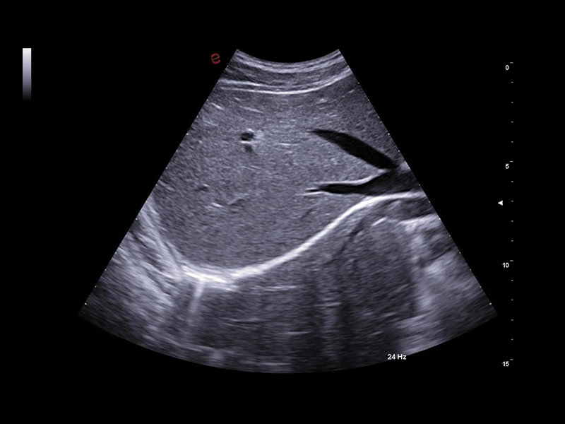
MyLab™A50 - Liver BMode
MyLab™A50 - Liver BMode
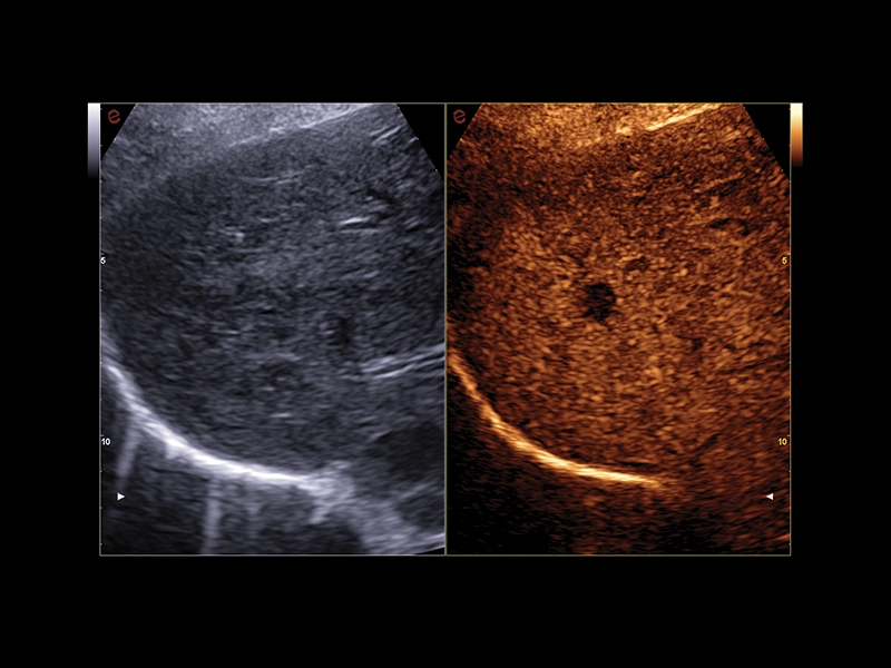
MyLab™A50 - Liver CnTi
MyLab™A50 - Liver CnTi
MyLab™A50 - microV kidney
MyLab™A50 - microV kidney
MyLab™A50 - MSK
MyLab™A50 - MSK
MyLab™A50 - PWD kidney
MyLab™A50 - PWD kidney
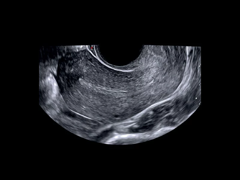
MyLab™A50 - Uterus
MyLab™A50 - Uterus
MyLab™A50 - vascular CFM
MyLab™A50 - vascular CFM
MyLab™A50 - vascular PW
MyLab™A50 - vascular PW
MyLab™A70 - 3D 4D
MyLab™A70 - 3D 4D
MyLab™A70 - AutoCM
MyLab™A70 - AutoCM
MyLab™A70 - AutoEF
MyLab™A70 - AutoEF
MyLab™A70 - Baby face
MyLab™A70 - Baby face
MyLab™A70 - BrightFlow
MyLab™A70 - BrightFlow
MyLab™A70 - Heart CFM
MyLab™A70 - Heart CFM
MyLab™A70 - Liver pathology
MyLab™A70 - Liver pathology
MyLab™A70 - Liver Bmode
MyLab™A70 - Liver Bmode
MyLab™A70 - MSK
MyLab™A70 - MSK
MyLab™A70 - QAI
MyLab™A70 - QAI
MyLab™A70 - XStrain RV
