Ultrasound Clinical Images
Just like space, some body parts are not visible to the naked eye: that’s why we need Esaote’s advanced diagnostic imaging technologies to be able to explore them and unlock their secrets.
Outstanding Ultrasound clinical images highlight the Esaote's focus in delivering innovative clinical solutions from prevention to therapeutic applications: a wide clinical gallery with high-res image quality means added diagnostic value and makes a difference for both clinicians and patients.
Today Esaote supports healthcare professionals with diagnostic imaging systems to deliver accuracy and precision of the clinical image as required by doctors on one hand, quality of diagnosis to improve the quality of life of the patients on the other and finally making what is essential visible to the eye and contributing to build a better future for everyone.
Filter by application
Filter by product
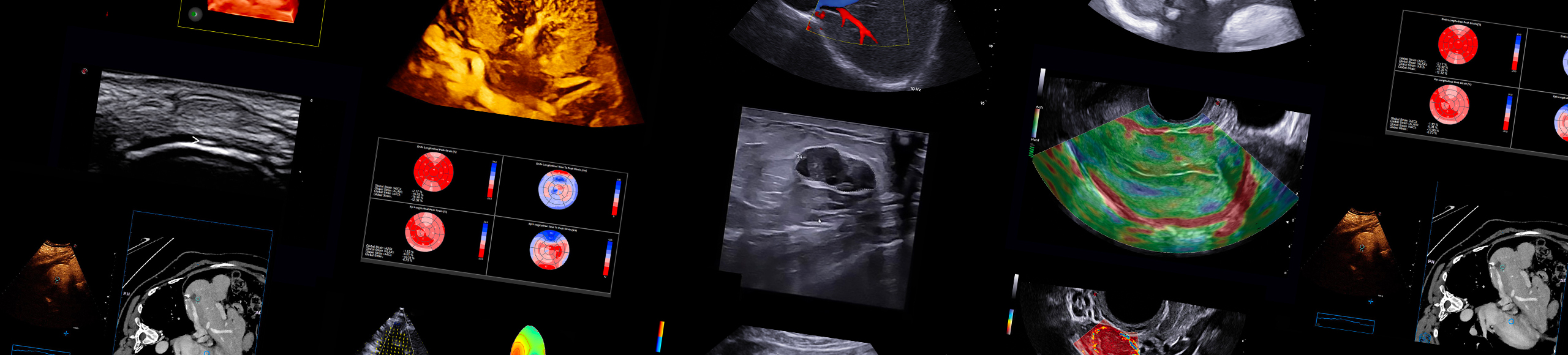
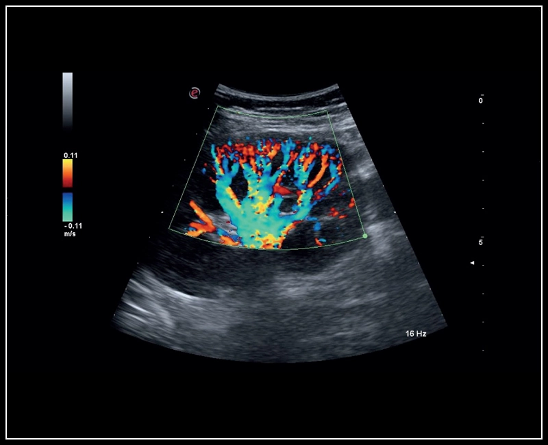
MyLab™Sigma - Fetal Willis circle with Power Doppler
MyLab™Sigma - Fetal Willis circle with Power Doppler
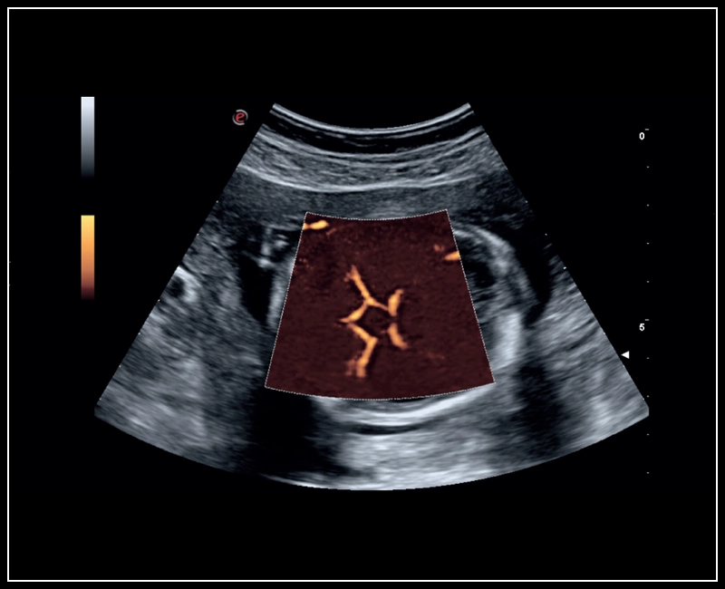
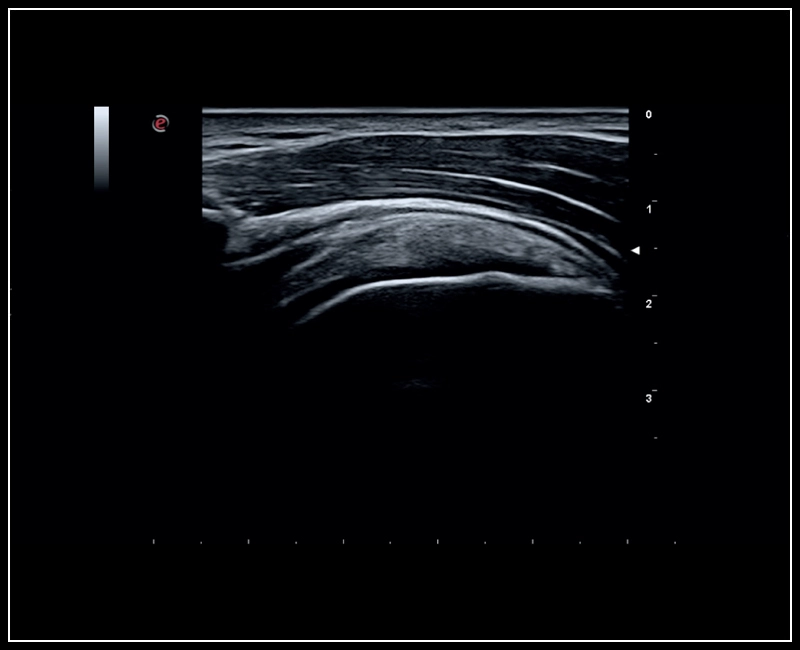
MyLab™Sigma - High Frequency Imaging B-mode of Intestine
MyLab™Sigma - High Frequency Imaging B-mode of Intestine
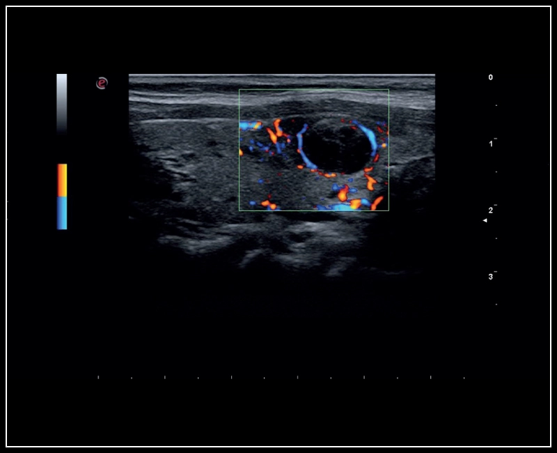
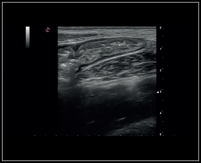
MyLab™Sigma - Thyroid lesion, imaging 2D with Color Doppler
MyLab™Sigma - Thyroid lesion, imaging 2D with Color Doppler
MyLab™Sigma - Kidney perfusion with high sensitivity Color Doppler mode
MyLab™Sigma - Kidney perfusion with high sensitivity Color Doppler mode
MyLab™Sigma - MSK imaging of the shoulder
MyLab™Sigma - MSK imaging of the shoulder
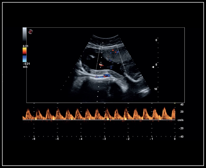
MyLab™Sigma - Umbelical cord PW Doppler mode
MyLab™Sigma - Umbelical cord PW Doppler mode
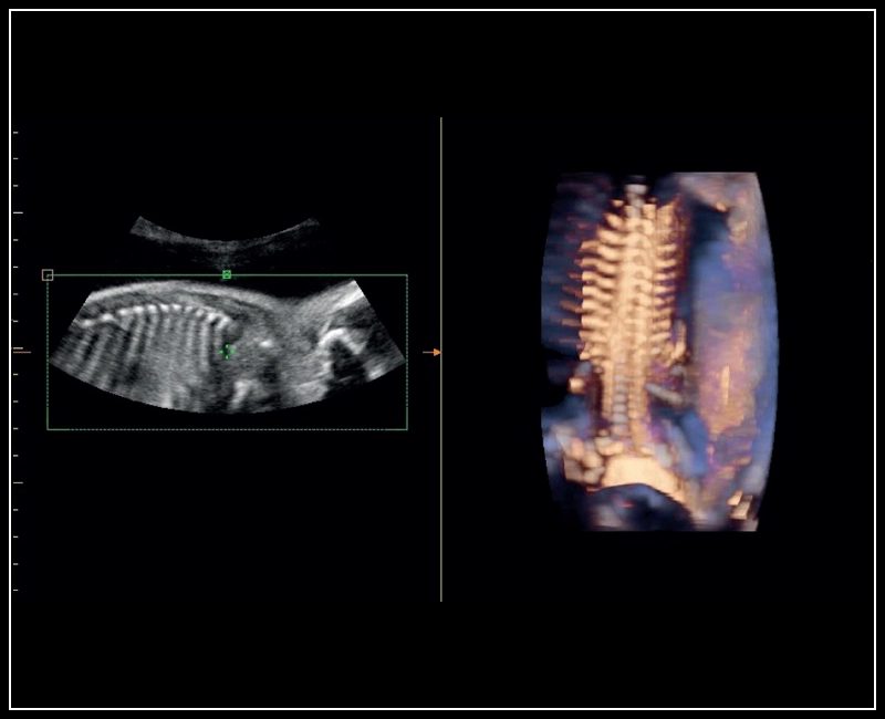
MyLab™Sigma - Semi transparent rendering of fetal spine
MyLab™Sigma - Semi transparent rendering of fetal spine
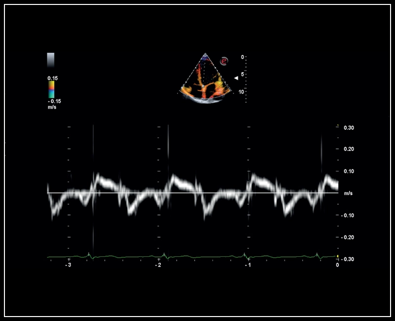
MyLab™Sigma - Mitral valve posterior leaflet analysis with Tissue Velocity Mapping
MyLab™Sigma - Mitral valve posterior leaflet analysis with Tissue Velocity Mapping
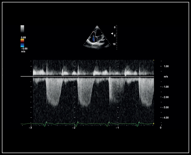
MyLab™Sigma - CW Doppler of Tricuspid regurgitation
MyLab™Sigma - CW Doppler of Tricuspid regurgitation
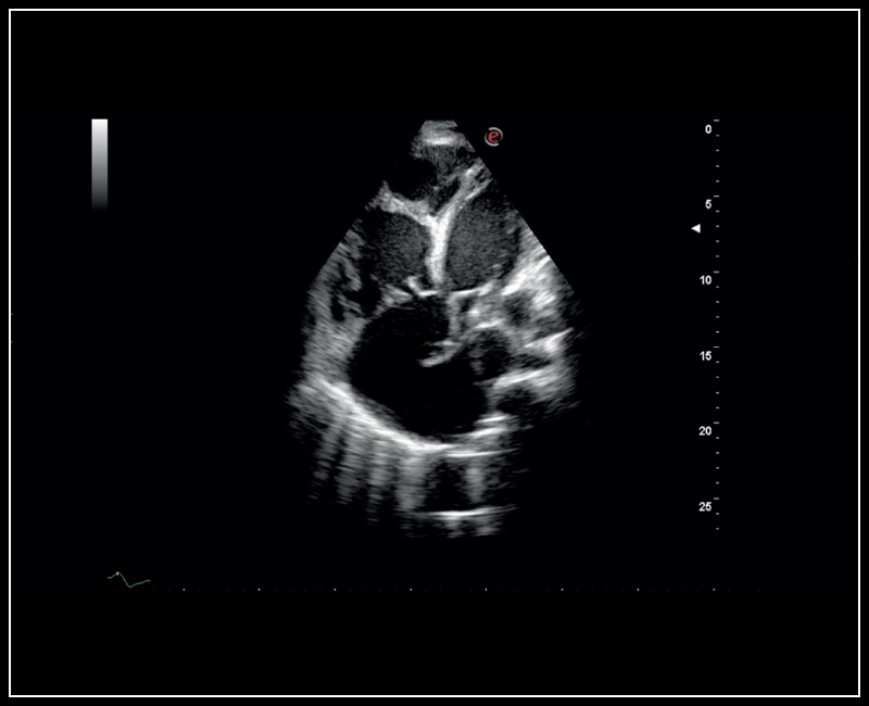
MyLab™Sigma - High definition B-mode cardiac imaging
MyLab™Sigma - High definition B-mode cardiac imaging
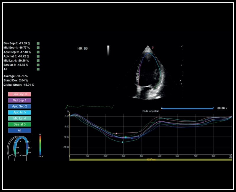
MyLab™Sigma - Left Ventricle XStrain 2D analysis
MyLab™Sigma - Left Ventricle XStrain 2D analysis
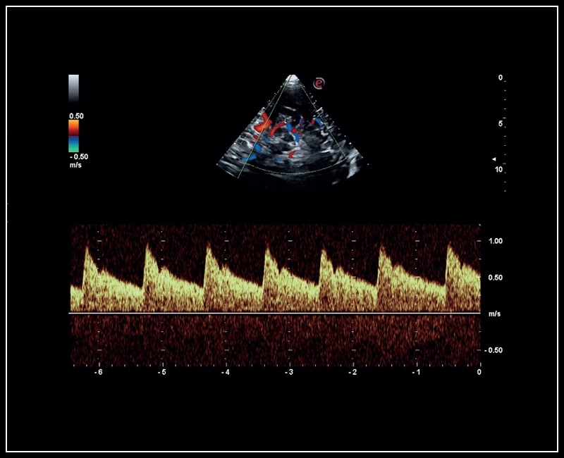
MyLab™Sigma - Mid cerebral artery investigation with PW Doppler mode
MyLab™Sigma - Mid cerebral artery investigation with PW Doppler mode
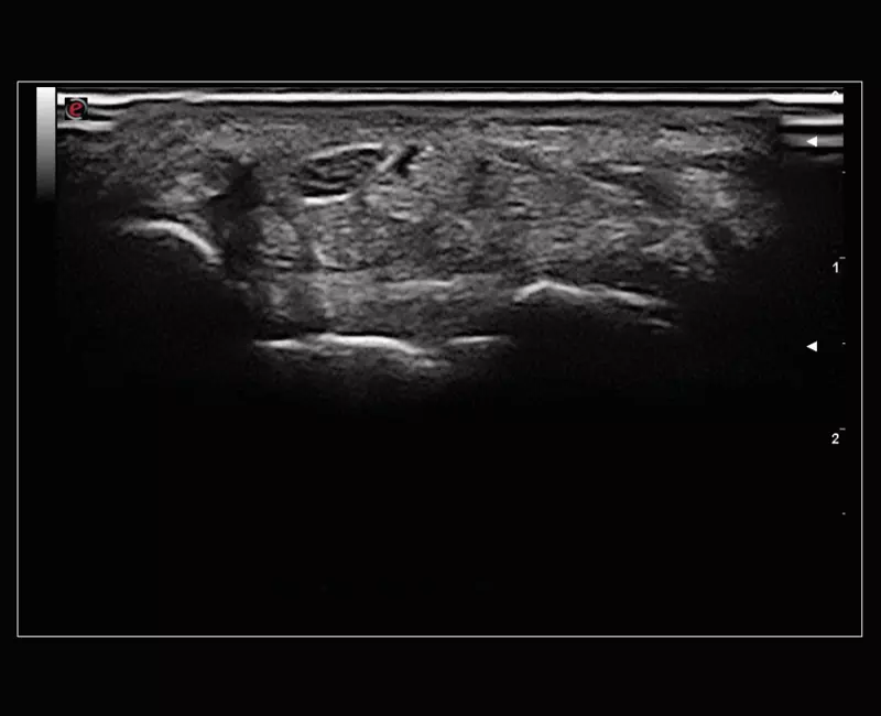
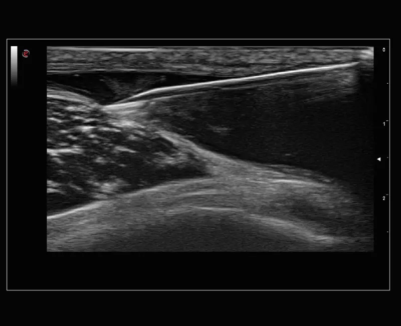
MyLab™Sigma Elite - B-Mode high resolution on median nerve
MyLab™Sigma Elite - B-Mode high resolution on median nerve
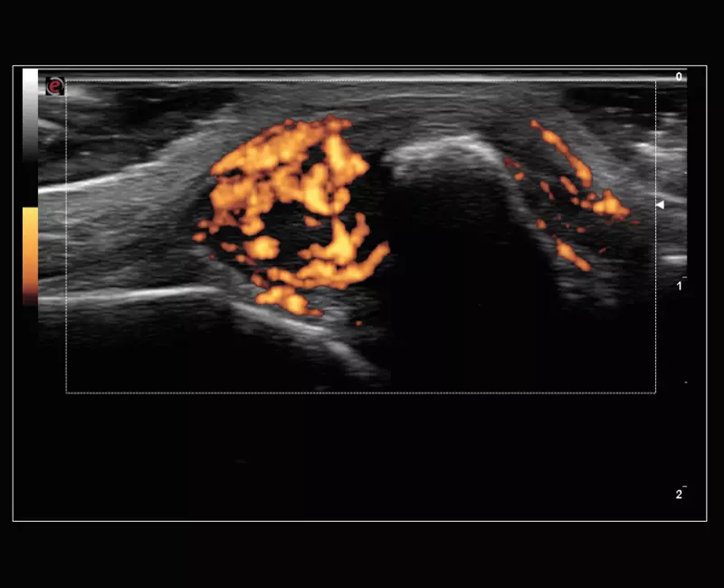
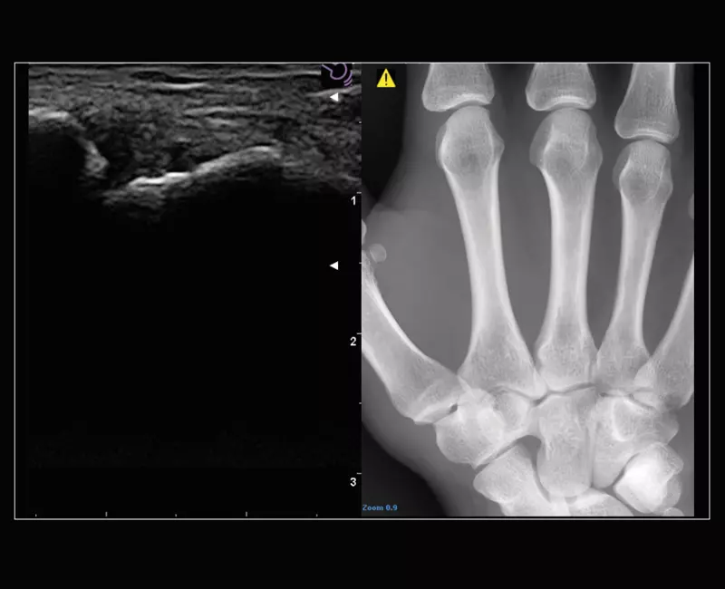
MyLab™Sigma Elite - Power Doppler on acute polyarthritis inflammation area with 18 MHz probe
MyLab™Sigma Elite - Power Doppler on acute polyarthritis inflammation area with 18 MHz probe
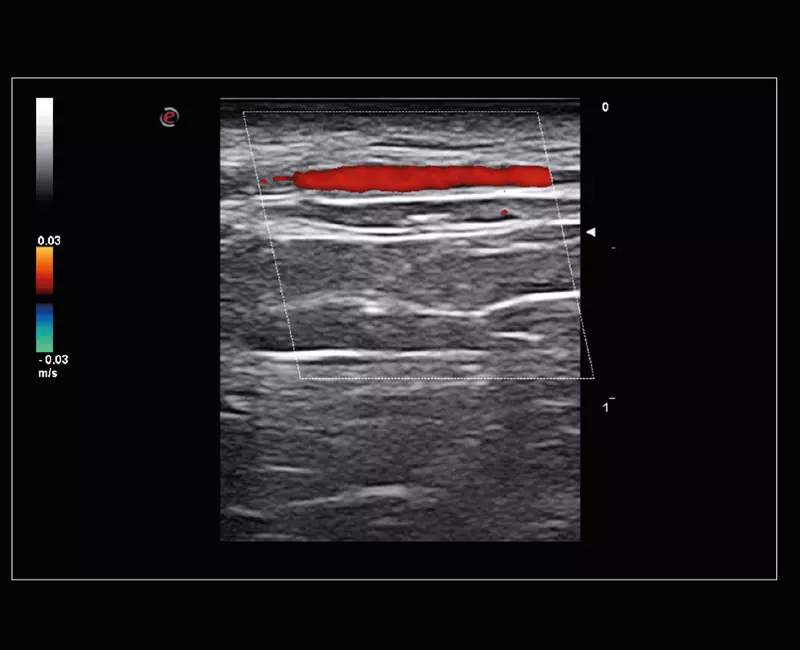
MyLab™Sigma Elite - Temporal Artery exploration with Very High Frequency probe (22 MHz)
MyLab™Sigma Elite - Temporal Artery exploration with Very High Frequency probe (22 MHz)
MyLab™Sigma Elite - Needle Enhancement for precise interventional procedures
MyLab™Sigma Elite - Needle Enhancement for precise interventional procedures
MyLab™Sigma Elite - Follow Up in real-time with a second modality
