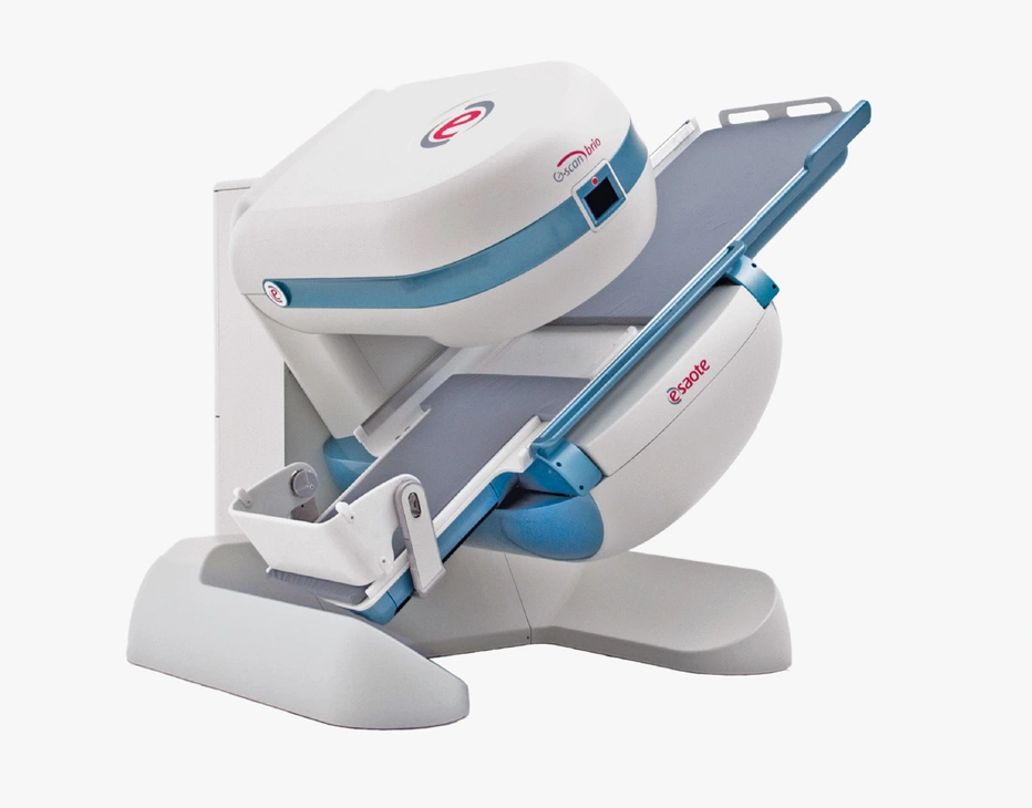
Weight-Bearing MRI
Esaote's revolutionary approach to Weight-bearing MRI set a new frontier in spine and knee MRI giving your clinical workflow the possibility of doing Weight-bearing MRI in a different way.
The open and tilting design is the new and innovative way of doing Weight-bearing MRI in which the position of the patient becomes an integral part of the outcome of the examination and allows patients to be imaged in the horizontal and vertical positions and to perform Weight-bearing MRI examinations viewing anatomy in orthostatic positions of daily activities than traditional supine positions.

- Provide more detail, better accuracy and greater confidence
Many symptoms and pathologies occur or are emphasized when the patient is in the weight-bearing position.
- Enhance the right biomechanics of several joints and anatomies
Esaote weight-bearing system ensures efficient diagnostic evaluations to reproduce and study joints in orthostatic position and find underlying pathologies.
- Patient-friendly atmosphere with Open MRI
Open MRI design fosters a patient-friendly setting, alleviating claustrophobia, simplifying positioning and promoting patient cooperation.
- Safety and Accessibility Focus
Low-field MRI enhances safety but also widens accessibility, making the MRI setting more comfortable and open to all people engaged in sports.
Traditional MRI exams are usually performed in supine positioning with the patient lying down on the scanner bed. However, this position does not reproduce the actual biomechanics of several joints and anatomies like knee, ankle and spinal columns which are subjected to weight-bearing stress when we are walking or simply standing up.
Therefore, Weight-bearing MRI can be a useful tool to reproduce and study these joints in orthostatic position and find underlying pathologies that are difficult be assess with the conventional supine MRI6.
INTERVIEWS
Testimonials

VIDEO
G-scan Spine Nevada revealing lumbar stenosis
Spine Nevada Minimally Invasive Spine Institute

VIDEO
The added value of G-scan
Glan Clwyd Hospital - Betsi Cadwaladr University Health Board - UK
Weight Bearing of the Spine
Chronic back pain is a common musculoskeletal issue and almost 50% of the population will develop Low Back Pain (LBP) in their life with a bigger incidence in middle-aged and older adults. LBP can affect the movement and the motion of those people and reflect the quality of their lives. It remains the leading global cause of years of life with disability (YLDs) worldwide and currently represents a social and economic burden1. MRI is one of the best image modalities to evaluate the integrity of spinal cords2. Nonetheless, the supine positioning of conventional MRI can, to some certain extent, alleviate the severity of the symptoms. On the other hand, the weight-bearing MRI can mimic the real physiologic conditions to give clinicians a better understanding of the anatomical relationship of the spine and surrounding tissues.


Doctor’s point of view
Compared with conventional supine MRI, the upright MRI provides a biomechanical approach, providing useful information to evaluate spinal stenosis, facet joint instability and spondylolisthesis4,5. MRI can also be an important instrument to evaluate how to proceed with spinal surgery and during the post-operative follow-up. Clinical study evidence that at least half of patients, who had spine surgery, had their treatment determined or modified after WB MRI6.

Q-Spine
In addition to the Weight Bearing functionality, Esaote supports the needs of the specialists with the Q-Spine software. Q-Spine is a support tool for the visualization and quantification of biomechanical changes of the Lumbar spine comparing supine and WB examinations.
Q-Spine functionality is based on the semiautomatic segmentation of the spine structures (vertebral bodies, spinal canal, foramina) both in Recumbent and Weight-Bearing: The reconstructed volumes are then used to perform automated measures providing numerical evidence of biomechanical modifications between Recumbent and Weight-Bearing positions, listed then in a pdf report to be eventually attached to image referral.
True-Motion
True-Motion can enable real-time visualization of musculoskeletal structures, with minimal time addition to routine exams. True-Motion could reveal intricate details that may go unnoticed in static images and could be particularly beneficial for post-operative evaluation. This is what makes True-Motion an invaluable specialized tool, ensuring unparalleled accuracy in diagnosis by meeting the unique diagnostic needs of patients. This innovation significantly expands the capabilities of medical imaging, being a key differentiation factor for certain pathologies in musculoskeletal examinations.
Metal Artifact Reduction
In our aging society, there is a growing prevalence of patients with orthopedic metal prostheses. This is evident in joint replacements, such as the knee or ankle, as well as in orthopedic implants used for intervertebral fusion, spine stabilization, and degenerative spine diseases. Consequently, the Metal Artifact Reduction (MAR) sequence is crucial in MRI examinations of patients with passive implants.

MAR employs an advanced gradients management system to effectively suppress or reduce in-plane metal distortions, ensuring optimal image quality. This innovation is particularly advantageous for post-operative patients, enhancing the imaging process and contributing to improved diagnostic precision. MAR uses an advanced gradients management system to effectively suppress or reduce in-plane metal distortions, ensuring optimal image quality. This innovation is particularly advantageous for post-operative patients, enhancing the imaging process and contributing to improved diagnostic precision in sports medicine.

Weight Bearing of lower extremity or limbs
The knee is one of the most complex joints and has a huge incidence of injuries that can be a source of long-term debility and social-economic impact7. Since most of the pathologies are posture-dependent and influenced by load, Weight Bearing MRI can provide added value tailored for the approach planning, especially for prosthetic ones. Knee exams performed with Weight-bearing MRI let the clinician gain more confidence issues on several pathologies such as Meniscal tears, ACL deficiencies and Patellofemoral displacement8. Also, WB can be beneficial to offer new insight to ankle and foot examination for the assessment of hallux valgus, midfoot instability, pes planus, fractures, and arthritic conditions of the hindfoot9.
Sources
- Fiani B, Griepp D W, Lee J, et al.; Weight-Bearing Magnetic Resonance Imaging as a Diagnostic Tool That Generates Biomechanical Changes in Spine Anatomy. Cureus 12(12): e12070
- Ge, L., Pereira, M.J., Yap, C.W. et al. Chronic low back pain and its impact on physical function, mental health, and health-related quality of life: a cross-sectional study in Singapore. Sci Rep 12, 20040 (2022).
- www.acr.org/-/media/ACR/Files/Practice-Parameters/mr-adult-spine.pdf
- Lang G, Vicari M, Siller A, Kubosch EJ, Hennig J, Südkamp NP, Izadpanah K, Kubosch D. Preoperative Assessment of Neural Elements in Lumbar Spinal Stenosis by Upright Magnetic Resonance Imaging: An Implication for Routine Practice? Cureus. 2018 Apr 6;10(4):e2440. doi: 10.7759/cureus.244
- Hebelka, H.; Rydberg, N.; Hutchins, J.; Lagerstrand, K.; Brisby, H. Axial Loading during MRI Induces Lumbar Foraminal Area Changes and Has the Potential to Improve Diagnostics of Nerve Root Compromise. J. Clin. Med. 2022, 11, 2122
- H.-S. Kim, G. Choi, W. H. Kim, H. J. Ma; Pohang K.R.;The effectiveness of the Weight-Bearing MRI in determining the optimal final surgical protocols for the patients with spinal disorders. European Society of Musculoskeletal Society of Radiology Congress 2018.
- Sam K. Yasen; Common knee injuries, diagnosis and management; Surgery (Oxford); Volume 41, Issue 4, 2023.
- Pages 215-222Bruno F, Barile A, Arrigoni F, Laporta A, Russo A, Carotti M, Splendiani A, Di Cesare E, Masciocchi C. Weight-bearing MRI of the knee: a review of advantages and limits. Acta Biomed. 2018 Jan 19;89(1-S):78-88
- Bruno F, Barile A, Arrigoni F, Laporta A, Russo A, Carotti M, Splendiani A, Di Cesare E, Masciocchi C. Weight-bearing MRI of the knee: a review of advantages and limits. Acta Biomed. 2018 Jan 19;89(1-S):78-88
Related system for Weight-bearing
Technology and features are system/configuration dependent. Specifications subject to change without notice. Information might refer to products or modalities not yet approved in all countries. Product images are for illustrative purposes only.
For further details, please contact your Esaote sales representative.

