How to design technology that saves lives: from the evolution of imaging to the I‑Genius case
The evolution of imaging over the last 30 years
In three decades, the way the human body is analysed through imaging has changed radically. In the early 1990s, the first dedicated MRIs were introduced: smaller, specialised solutions designed specifically to study the extremities of the body, such as knees, hands, feet, and ankles. The aim was practical: to reduce the workload of large hospital MRI systems, which were often operating at full capacity, by providing a more agile alternative for those examinations that would otherwise remain on the waiting list. These systems also offered a high level of patient comfort: patients could sit comfortably in a chair, insert only the limb to be examined into a small opening, and read or relax, almost forgetting they were undergoing a medical procedure.
In 1992, Esaote was the first company to introduce an open MRI system specifically designed for joint imaging. This pioneering solution also addressed the need to make the examination accessible to claustrophobic people, children, and overweight patients, overcoming the limitations of traditional closed tunnel systems.
The following decade marked a gradual yet significant expansion of clinical applications. Imaging capabilities were extended from the shoulder to the different sections of the spine, lumbar, dorsal, and cervical, and finally the study of the head. During this period, MRI systems became increasingly versatile while maintaining their open architecture and the use of permanent magnets. This evolution also led to the development of innovative solutions for spine imaging in the upright position, introducing new diagnostic perspectives and expanding clinical insight.
For Esaote, 2021 marked the introduction of the first low-field total body MRI systems. The key challenge was to ensure high diagnostic image quality while adopting a more compact, environmentally sustainable, and cost-effective technological approach.
Today, an even more fascinating new chapter is unfolding. Imaging technologies are evolving beyond their traditional diagnostic role towards intraoperative applications, enabling their use directly during surgical procedures and opening up new opportunities for clinical practice.
The three priorities: image, usability, comfort
What drives the design of a new imaging system today? According to Fabrizio Ferrando, MRI R&D Director at Esaote Spa, the hierarchy of priorities is clear. "Image quality comes first, because the physician’s diagnosis is based on the image itself. Next is ease of use: operators must be able to work under optimal conditions, achieving consistent, standardised results through an intuitive interaction with the system”.
Automation has also become an increasingly central element in system design: "Solutions are being developed to automate certain phases of the workflow, including with the use of artificial intelligence. Take MRI: it is a solution that has many parameters that can be changed. It is like having a professional manual camera, with plenty of opportunities for customisation as required. Today, our goal is to develop solutions that allow healthcare professionals to achieve high-quality results without having to manage unnecessary operational complexity”.
Patient comfort represents the third key pillar - an aspect well known to anyone who has undergone an MRI examination in a closed system. To take up the metaphor, MRI can be compared to long-exposure photography. As Ferrando explains: "If the patient does not stand still, the image is moved. And remaining still for half an hour is not always easy. When the patient is comfortable, movement is reduced, and image quality improves accordingly”.
In closed, tube systems, this issue is further amplified. Depending on the anatomical area being examined, patients are often required to maintain uncomfortable or unnatural positions, which can negatively affect both the examination experience and the final diagnostic outcome.
When clinical outcomes become the primary goal: the I‑Genius case
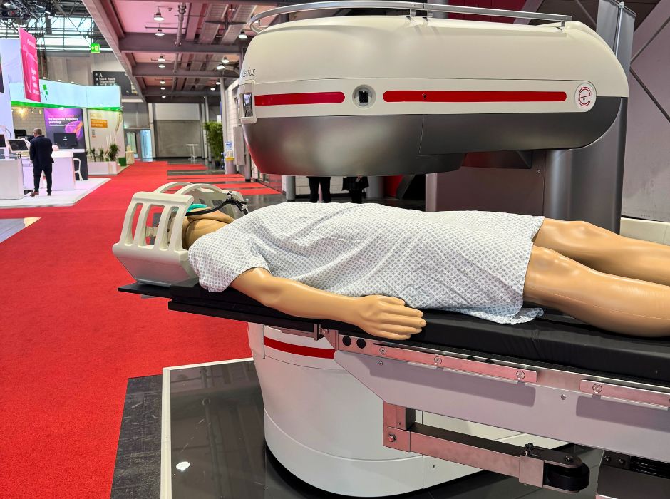
But what happens when the clinical context reshapes the hierarchy of design priorities? This is precisely the scenario that led to the development of I‑Genius, the latest product launched by Esaote, an MRI system designed to operate inside the operating theatre during brain surgery, particularly in the treatment of gliomas.
"In this case, patient comfort is no longer the primary concern”, explains Ferrando, “since the patient is under general anaesthesia with the cranial cavity open. Here, imaging plays a critical role in supporting the neurosurgeon in achieving complete tumour resection". The clinical stakes are extremely high: " The leading cause of tumour recurrence in gliomas is incomplete removal of the neoplastic tissue”.
The problem to be solved: one hour per control
The technique, known as intraoperative MRI, consists of alternating surgery and imaging phases. “In this context, imaging is not for diagnostic purposes, because the diagnosis has already been established” Ferrando clarifies, “but it allows the surgeon to assess how much tumour tissue has been removed and what remains. And it also helps to find the best route to the tumour, making its way into the brain tissue in the least damaging way possible”.
With conventional high-field MRI systems, 1.5 or 3 Tesla superconducting magnets, performing an intraoperative check is a logistical challenge. The patient must be transported from the operating room to the scanner and then back. "It takes about an hour," says Ferrando. " For a surgery that already lasts several hours, with the patient under anaesthesia, I can only perform a single check, usually at the end of the procedure”.
" With a system that allows multiple intraoperative checks—potentially more than five during a single operation—I can leverage real-time imaging to confidently remove the entire tumour, without significantly extending anaesthesia time. This capability enhances surgical precision and improves clinical outcomes”.
The intuition: the rotating bed
The idea behind I-Genius exploits a technical feature of low-field MRI systems: the reduced stray magnetic field. Permanent magnets generate a much weaker field than superconducting systems, which creates new possibilities for intraoperative use. "This enables us to keep the patient stationary while the MRI bed itself is able to rotate and slide in and out of the magnet cavity, guided precisely by a compass system as needed for either surgery or imaging", Ferrando explains.
In practice, the patient never moves. The bed rotates (compasses) from the operating position within the magnet, allowing images to be captured, and then returns to the surgical position. "It’s an operation that can be performed by a single operator in just a few minutes”, Ferrando adds.
Planning: stepping out of the comfort zone and looking for strategic partners
Designing a system like I-Genius meant facing completely new challenges. "We’re not talking about the technological aspects we normally handle at Esaote", Ferrando admits. "Here we had to deal with issues somewhat outside our usual design domain.
The first challenge was the surgical table. "It is a highly complex component because it must move in multiple directions: it rotates, it rises, it moves horizontally and vertically. Integrating an MRI and a surgical bed is not a trivial matter”. To address this, Esaote partnered with an Italian company specialised in the design and production of surgical tables. "We realised that in order to solve this problem, we had to collaborate with those who had the expertise on the beds. Attempting this alone would have slowed us down and distracted us from our core goal.
Next came the challenge of material compatibility. Everything that enters the MRI system must be MRI-safe, meaning it must not interfere with the magnetic field or generate imaging artefacts. Metals in general are problematic, even non-ferromagnetic ones, such as aluminium, which has a weak magnetic susceptibility but, if present within the device cavity, leads to the generation of eddy currents that introduce artefacts into the image. Lightweight, strong materials like carbon fibre, commonly used in surgical beds for C-arm X-ray systems, are not compatible with MR imaging.
The solution was a composite multilayer material made of paper, wood fibre and resin, rigid and resistant: "It is completely invisible to the MRI, it does not introduce any artefacts, but provides the necessary robustness to support the patient even when the surgical bed is in a highly overhanging position”.
The same challenge arose for the cranial stabiliser, which secures the patient's head. "On the market, these components are typically made of steel. We switched to a plastic material, with a greater thickness to guarantee the necessary tightness, rigidity and mechanical stability in the face of all the pressure actions that the surgeon performs on the cranial box during surgery”.
Even the coil required a complete rethink: "It needed to be large enough to accommodate the cranial stabiliser, but increasing its size reduces signal efficiency. Designing it required careful compromise to balance coverage and performance”.
The field test: twenty-eight interventions
The validation was validated in Argentina. Dr Roberto Rafael Herrera, Chief of Neurosurgery at Clínica Adventista Belgrano in Buenos Aires, became the key opinion leader in the product’s development. The results, published in 2024 in Medical Research Archives, are notable: among 28 patients undergoing cerebral glioma surgery with the low-field system, total gross resection (greater than 99%) was achieved in 82% of cases. The average imaging session lasted only 12 minutes. And most importantly, in 93% of cases, the first intraoperative scan revealed residual tumour tissue, leading to further resections that would otherwise not have been performed.
The ability to perform repeated scans, ranging from one to four per operation, enabled more complete tumour removal without increasing complications: 85.7% of patients experienced no new neurological deficits following surgery.
Designing for results
The story of I-Genius tells something important about how medical technology is designed today. You don't start with the machine, you start with the clinical problem. Solving that problem often requires stepping beyond your traditional areas of expertise, seeking strategic partnerships, experimenting with new materials, and accepting necessary compromises.
"The goal was to deliver a complete solution that enables an optimised workflow and high-quality imaging to monitor tumour resection", Ferrando concludes. "A solution that truly adds value from the perspective of surgical outcomes".
Over the past thirty years, technology has evolved dramatically. But the guiding spirit remains the same: responding to real clinical needs.
The study
Herrera RR et al, Intraoperative Magnetic Resonance Imaging in Brain Glioma Surgery Using Low-field system. Presentation of the First Twenty-eight Procedures, Medical Research Archives, 2024; 12(6). DOI: 10.18103/mra.v12i6.5387
This article is part of the editorial project “Technology and empathy: the new story of care with Esaote and PEOPLE”, which is a collaboration between Esaote and PEOPLE Magazine.
Read other articles

MAGAZINE
Behind the scenes of the medical infrastructure supporting a major sporting event: what it takes to ensure care at the Winter Olympics
The organisation of imaging and healthcare in a multi-venue event such…

CARE
Caring for the carer: a still underestimated medical responsibility
Behind every person facing a serious illness, there are others who…
CARE
Prevention begins within the company: breast cancer screening in the workplace
October marks Breast Cancer Prevention Month - a time to spotlight a…

CARE
Technology and empathy: the new narrative of care with Esaote and PERSONE
We are living in an extraordinary age for medicine: we’ve never had…
CARE
How MRI technology enables democratising access to diagnostics in remote areas
A new open MRI scanner brings advanced diagnostics directly to people.…
